研究问题与核心发现
ZFP36L2 帮助细胞清除应激相关 RNA,恢复 LGR5+ 肠干细胞状态。 缺失它会损害肠道修复并削弱癌细胞早期定植,同时使幸存癌细胞出现非经典分化状态(PDF 第 3–10 页)。
研究将正常组织再生与癌细胞可塑性放在同一框架中:应激反应启动之后,细胞如何退出这一程序、重新进入干细胞状态?
研究设计
| 层次 | 模型与技术 | 解决的问题 |
|---|---|---|
| 细胞状态 | 正常与肿瘤上皮单细胞数据 | ZFP36L2 与干细胞及应激程序的联系 |
| 肠道再生 | 上皮特异性敲除、DSS 损伤、Lgr5-DTR 消融、类器官 | ZFP36L2 是否参与损伤后去分化 |
| 转移与分化 | 患者来源类器官、原位与肝定植模型 | 缺失后的生长、定植及命运变化 |
| RNA 调控 | 活细胞成像、FRAP、HyperTRIBE、ActD、SLAM-seq | 凝聚体动态、RNA 结合与衰减 |
实验设计:PDF 第 2–9、12–18 页。
发现一:损伤后的干细胞状态恢复受损
在 DSS 损伤恢复和 LGR5+ 细胞消融模型中,Zfp36l2 缺失均影响上皮再生。类器官实验进一步将这一表型与细胞内在的去分化能力联系起来(PDF 第 3–4 页,图 2)。
这组实验把 ZFP36L2 从细胞状态相关因子,推进到损伤后再生过程中的功能调控因子。
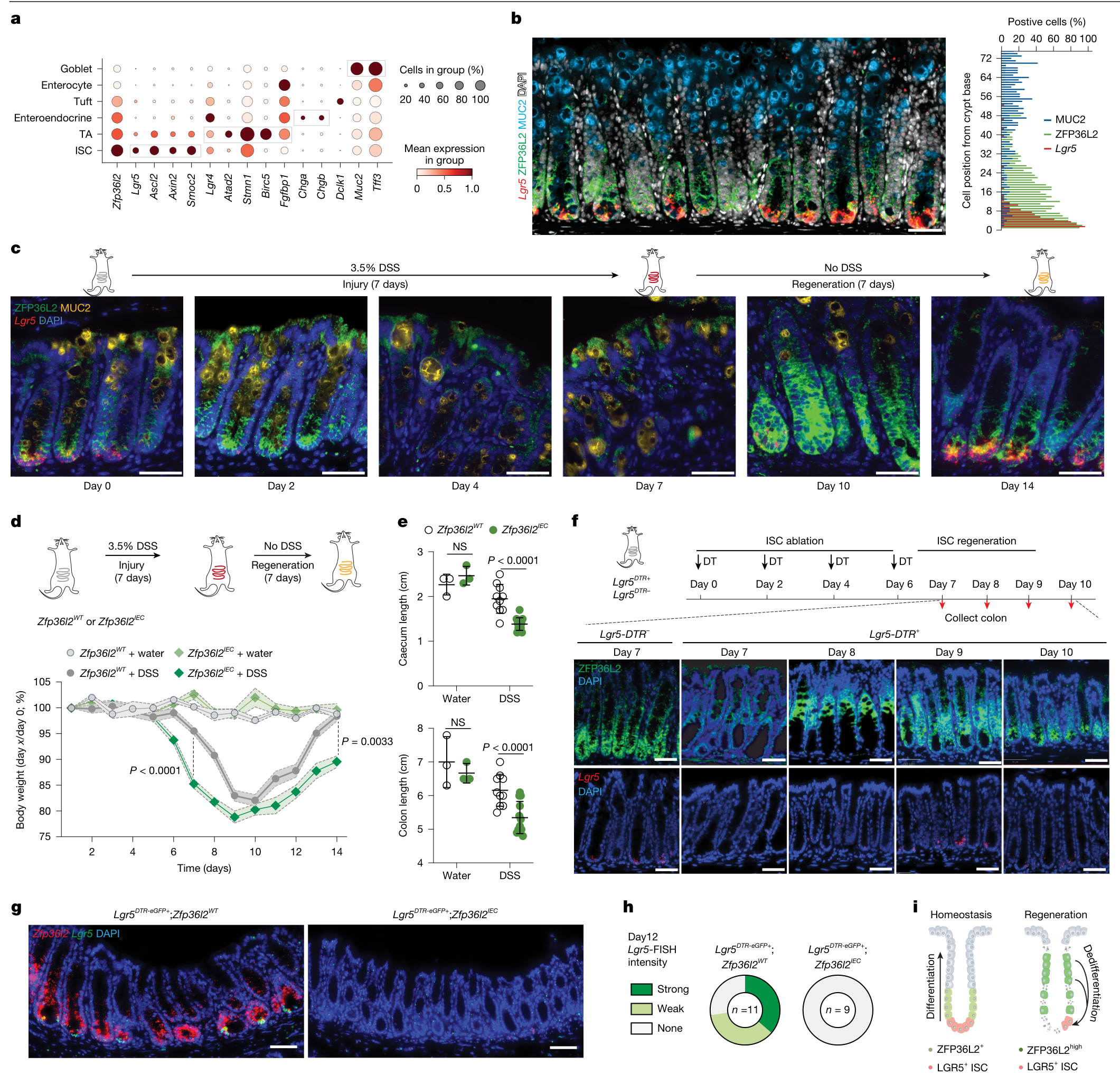
图 2 · 损伤修复与去分化。原文 PDF 第 4 页。
发现二:转移定植减少,分化路径发生改变
ZFP36L2 扰动降低了部分模型中的肝转移定植,但幸存细胞仍可继续生长,并出现 CHGB 等非经典分化相关变化(PDF 第 5–7 页,图 3)。
定植能力与后续细胞命运是两个不同的结果。 抑制经典干细胞状态恢复的同时,癌细胞也可能转向其他分化路径。
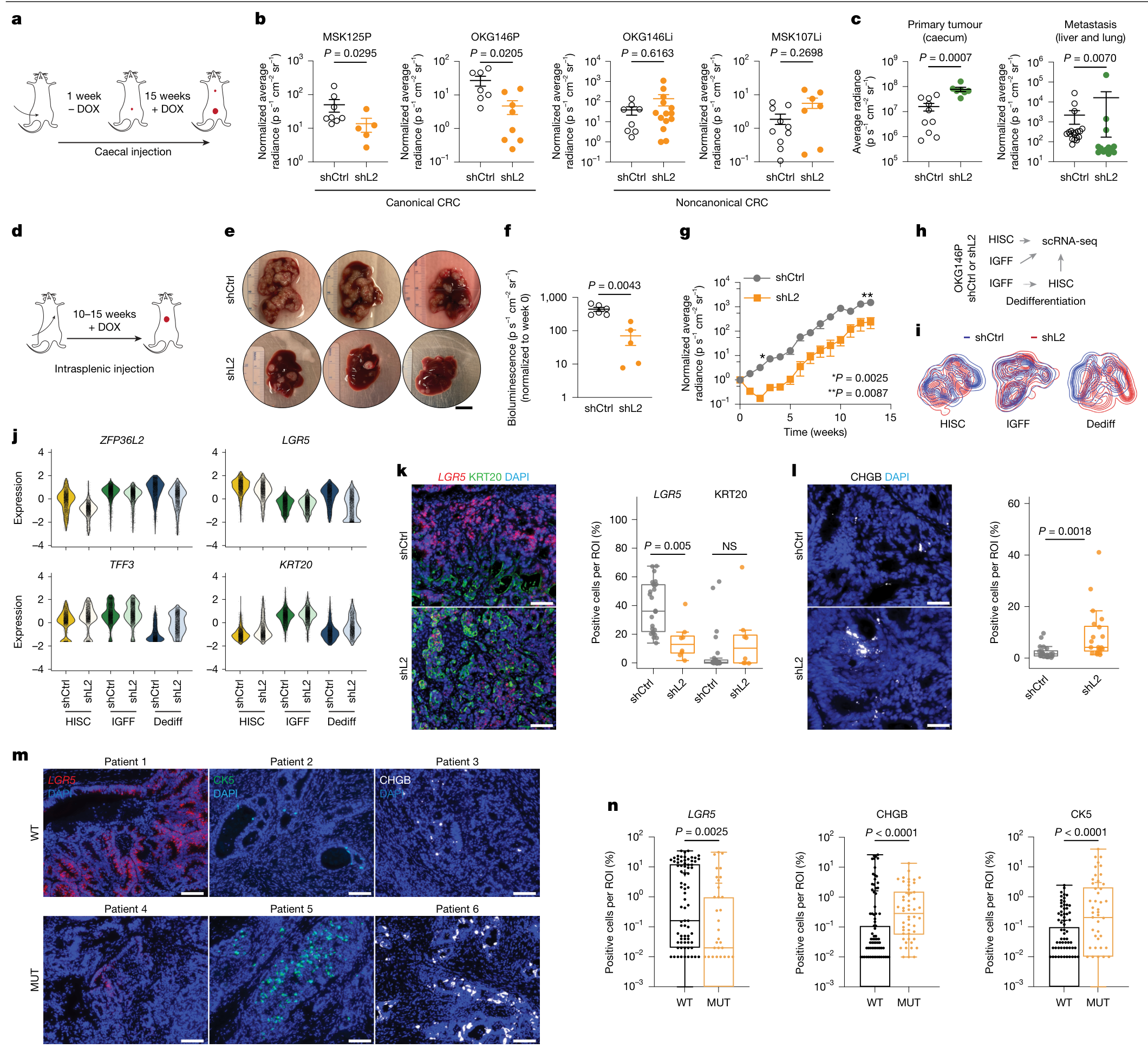
图 3 · 转移定植与非经典分化。原文 PDF 第 6 页。
机制:凝聚体动态与应激 RNA 衰减
活细胞成像、FRAP 和 RNase 实验显示,ZFP36L2 相关凝聚体具有动态性和 RNA 依赖性(PDF 第 7–8 页,图 4)。
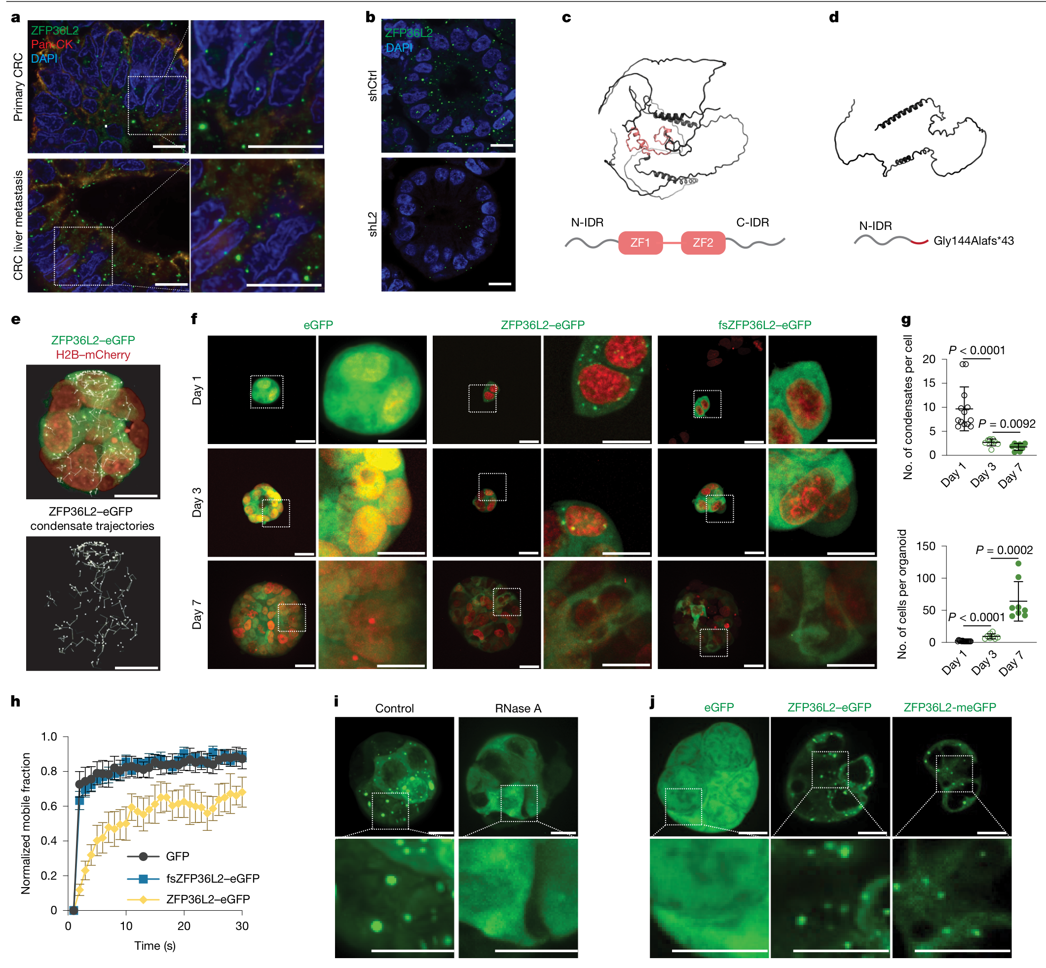
图 4 · RNA 依赖的动态凝聚体。原文 PDF 第 8 页。
HyperTRIBE 定位 RNA 相互作用,ActD 与 SLAM-seq 检测 RNA 稳定性。结合 ZFP36L2 扰动后的变化,结果支持它参与应激相关转录本的降解,推动细胞退出应激状态(PDF 第 9 页,图 5)。
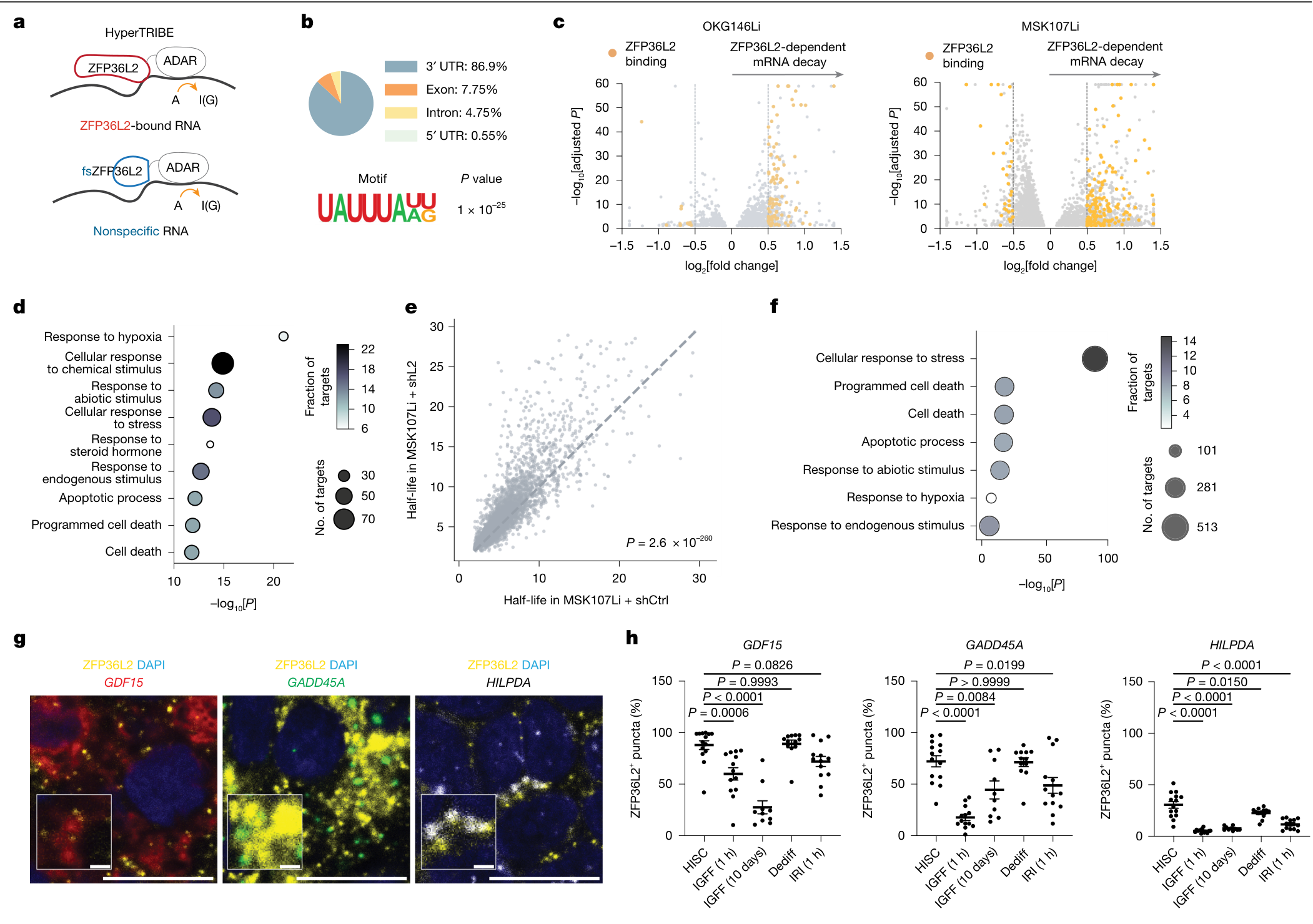
图 5 · RNA 结合与衰减。原文 PDF 第 9 页。
这项研究带来的新线索
应激退出也是命运调控的一环。 RNA 降解将短期应激响应与后续干细胞状态恢复联系起来,为理解再生时序提供了分子线索。
正常再生与癌症可塑性共享部分程序。 同一个调控因子参与损伤修复与转移定植,干预效果也随组织和细胞状态变化。
癌细胞命运具有替代路径。 经典干细胞程序受限后出现的非经典分化,是理解转移适应与干预后细胞状态演变的重要方向(PDF 第 10 页)。
可延伸阅读:Moorman 等,Progressive plasticity during colorectal cancer metastasis,Nature(2025);本文 ref. 10。
主图与表格 · 5 张
Fig. 1 · PDF 第 2 页
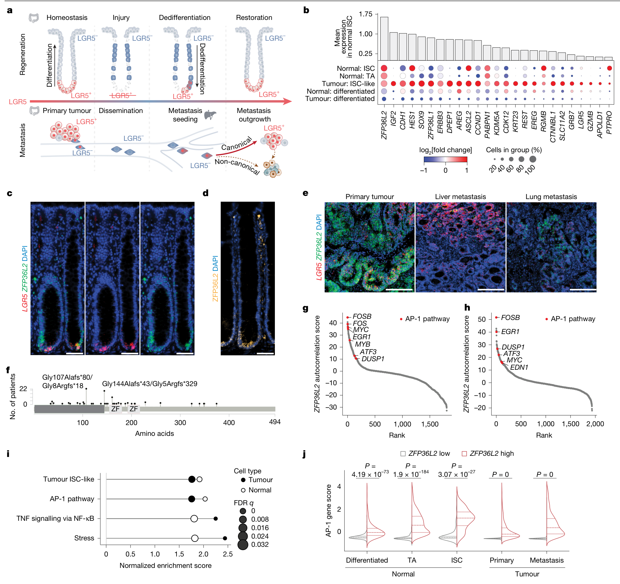
Fig. 1 | ZFP36L2 is expressed by ISCs and is associated with the AP-1 gene program. a, Schematic of cell-state plasticity and dynamics during intestinal regeneration (top) and CRC (bottom). b, Dot plot of tumour ISC-like module10 gene expression in normal colon (Normal) and CRC (Tumour) epithelial cells from 25 patients. The dot size indicates the per cent cells (rows) expressing each gene (columns). Colour scale indicates the log2 fold change in average gene expression. Bar plots show the mean expression of each gene across normal colon ISCs. Genes meeting false discovery rate (FDR) < 0.05 using the Wilcoxon rank-sum test (ISC versus all other epithelial subtypes) are shown. c,d, Representative LGR5 and ZFP36L2 RNA FISH (c) and ZFP36L2 IF (d) images of human normal colon crypts. e, Representative LGR5 and ZFP36L2 RNA FISH images of human primary CRC tumours (n = 2), liver metastases (n = 2) and lung metastases (n = 2) from 2 donors. f, Lollipop plot showing ZFP36L2 mutation frequency in the TCGA63, DFCI64 and MSKCC whole-exome sequencing CRC cohorts. n = 96 tumours. g,h, ZFP36L2 autocorrelated gene programs in normal colon (g) and CRC epithelial cells (h). Red, top AP-1 family genes65. n = 12,955 normal colon (24 donors) and 25,317 CRC cells (25 patients). FDR < 0.05. i, GSEA of genes (Supplementary Table 1b–f) ranked by positive ZFP36L2 autocorrelation (Methods), which revealed conserved pathways enriched in normal (white dots) and tumour (black dots) epithelial cells. n = 12,955 normal colon (24 donors) and 25,317 CRC cells (25 patients). FDR < 0.05. j, Violin plots showing AP-1 pathway gene expression in ZFP36L2-high and ZFP36L2-low populations (top and bottom 10 percentile expression). n = 1,806 differentiated, n = 1,326 TA and n = 230 ISC cells from normal colons (24 donors) and 2,622 primary CRC and 2,444 CRC metastasis cells (25 patients). Two-sided Mann–Whitney U-test. Scale bars, 50 µm (c,d) or 200 µm (e).
Fig. 2 · PDF 第 4 页
Fig. 2 | Zfp36l2 is required for injury-induced dedifferentiation during intestinal regeneration. a, Zfp36l2 expression across epithelial cells, as measured by scRNA-seq of colonic crypts (21,000 cells from 11 mice)66. Marker genes for each cell type are indicated by boxes. b, Representative images (left) and quantification (right) of Lgr5 RNA FISH and ZFP36L2 and MUC2 IF in colon tissue from 7-week-old C57BL/6J mice based on the cell position along the crypt base (position 0) to lumen axis. n = 2,275 cells from 31 crypts. c, Representative images of Lgr5 RNA FISH and ZFP36L2 and MUC2 IF in colon tissue collected at the indicated time points from mice treated with 3.5% DSS for 7 days followed by recovery for 7 days. n = 3 mice per time point. Expanded fields of view are provided in Supplementary Figs. 1 and 3. d, Top, schematic of the experiment. Bottom, daily body weight measurements. n = 5 (water) mice; n = 10 (wild-type (WT)) and 14 (IEC) (DSS-treated) mice. Mean (solid line; dots, individual time points) ± s.e.m. (shaded area; dashed lines, s.e.m. boundaries); two-tailed Mann–Whitney U-tests at day 7 and day 14. e, Caecum (top) and colon (bottom) lengths on day 14. n = 3 mice (water); n = 10 (WT) and 12 (IEC) (DSS-treated) mice. Mean ± s.d.; two-tailed Mann–Whitney U-test. NS, not significant. f, Lgr5DTR ablation. Top, schematic of the experiment. Bottom, representative images (n = 2 mice) of colon sections stained by Lgr5 RNA FISH or ZFP36L2 IF at the indicated time points. Expanded fields of view are provided in Supplementary Fig. 5. Controls: Lgr5DTR-negative mice treated with 4 doses of DT over 7 days. g,h, Mice (8–10 weeks old) were administered DT every 2 days for 4 doses. Mice were euthanized and colons were collected 12 days after the first DT dose. g, Images of mouse Lgr5 and Zfp36l2 mRNA FISH on day-12 Swiss-roll colon sections. h, The number of mice showing regeneration of the Lgr5 FISH signal. i, Schematic of the expression and function of ZFP36L2 in injury-induced dedifferentiation to drive regeneration in the mouse colon. Scale bars, 50 µm (b,c,f,g).
Fig. 3 · PDF 第 6 页
Fig. 3 | Loss of ZFP36L2 inhibits CRC metastasis seeding and ISC dedifferentiation but promotes non-canonical differentiation. a–c, Orthotopic caecal xenotransplantation experiments. a, Schematic of the experiment. DOX, doxycycline. b, In vivo abdominal BLI average radiance, normalized to BLI at the time of doxycycline diet initiation. shL2, shZFP36L2. n (left to right) = 8, 5, 7, 8, 8, 14, 10 and 7 animals per group. Mean ± s.e.m.; two- tailed Mann–Whitney U-tests. c, End point ex vivo BLI of MSK107Li samples. Metastasis signals were normalized to orthotopic caecal signals in the same animals. n = 10 (shCtrl) and 7 (shL2) mice. Mean ± s.e.m.; two-tailed Mann– Whitney U-tests. d–g, Orthotopic liver metastasis seeding experiments. d, Schematic of the experiment. e, Representative ex vivo images of MSK107Li liver metastases. f, Average radiance of week 13 metastasis normalized to week 0 BLI. n = 6 (shCtrl) and 5 (shL2) mice. Mean ± s.e.m.; two-tailed Mann–Whitney U-tests. g, Weekly whole-body in vivo liver BLI (mean ± s.e.m.) normalized to week 0. n = 7 (shCtrl) and 5 (shL2) mice. Two-tailed Mann–Whitney U-tests. h, Schematic of the experiment. i. Kernel density estimate contour plots of scRNA-seq data from h, showing the overlap and divergence of cell states in OKG146P organoids transduced with shCtrl or shL2 and cultured in HISC, IGFF or dedifferentiated (Dediff) conditions. j, Violin plots showing expression of the indicated markers. k,l, Representative immunostaining (left) and quantification (right) of LGR5 RNA FISH and KRT20 IF in MSK107Li (k; see also d–g) and CHGB IF in OKG146Li-MS2 liver metastasis (l). The graphs show the per cent LGR5 and KRT20+ cells in each of the 26 regions of interest (ROIs) (252,330 cells; shCtrl) and 8 ROIs (194,530 cells; shL2) or CHGB+ cells in each of the 21 ROIs (254,403 cells; shCtrl) and 20 ROIs (66,338 cells; shL2) from 3 mice per group. Box plots show the interquartile range, with the line indicating the median, and whiskers the minimum and maximum values. Two-tailed Mann– Whitney U-tests. m,n, Representative images (m) and quantification (n) of LGR5 RNA FISH and CK5 and CHGB IF in WT or ZFP36L2 mutated (MUT) paired primary–metastasis CRC samples from patients. n = 8 (ZFP36L2 WT) and 4 (ZFP36L2 MUT) samples. LGR5 RNA FISH: 91 ROIs (1,301,960 cells; ZFP36L2 WT) and 43 ROIs (1,154,616 cells; ZFP36L2 MUT). CHGB and CK5 IF: 113 ROIs (1,814,441 cells; ZFP36L2 WT) and 50 ROIs (1,302,471 cells; ZFP36L2 MUT). Box plots show the interquartile range, with the line indicating the median, and whiskers the minimum and maximum values. Two-tailed Mann–Whitney U-tests. Scale bars, 50 µm (l), 100 µm (k,m) or 1 cm (e).
Fig. 4 · PDF 第 8 页
Fig. 4 | ZFP36L2 forms RNA-dependent, stress-responsive biomolecular condensates. a, Representative endogenous ZFP36L2 and pan-cytokeratin (Pan-CK) IF images of primary CRC and CRC liver metastasis from patient KG146. b, ZFP36L2 IF in MSK107Li organoids transduced with shCtrl (top) or shZFP36L2, demonstrating efficient knockdown and staining specificity. c,d, Top, AlphaFold2-predicted protein structure of human WT ZFP36L2 (c) and CRC patient hotspot mutation Gly144Alafs*43 (d). Bottom, schematic of the protein structures indicating the CCCH zinc fingers (ZFs) and the N-terminal and C-terminal IDRs. Red, ZF domains (amino acids 153–219); grey, IDRs. e, Representative images of maximum-intensity projection of motion-artefact- corrected 3D time-lapse video of OKG146Li organoids co-expressing ZFP36L2– eGFP and H2B–mCherry. Top, condensate positions and trajectories overlaid on the original image. Bottom, corresponding trajectory rendering shown separately. f, Representative images of single z plane confocal live-cell microscopy of MSK107Li H2B–mCherry (red) organoids with doxycycline- inducible expression of eGFP, WT ZFP36L2–eGFP or fsZFP36L2–eGFP. Organoids were plated as single cells on day 0, and doxycycline was added 24 h before imaging. g, Mean number of condensates per cell (top) and number of cells per organoid (bottom) at the indicated time points after single-cell seeding of ZFP36L2–eGFP-expressing MSK107Li H2B–mCherry organoids. Data are the mean ± s.d., and data points represent individual organoids (n = 8). Two-tailed Mann–Whitney U-tests. h, Normalized fluorescence intensity of MSK107Li organoids after photobleaching. Data representative of four replicates (mean ± s.d.). i, Live-cell imaging of ZFP36L2–eGFP-expressing MSK107Li organoids, treated with or without 1 μg μl–1 RNase A, 30 min after treatment. j, Live-cell imaging of OKG146Li organoids expressing doxycycline- inducible eGFP, ZFP36L2–eGFP or ZFP36L2–meGFP. Day 3 organoids were treated with 2 μg ml−1 doxycycline for 24 h before imaging. Insets are shown at higher magnification (bottom). ZFP36L2–meGFP exhibited condensate formation comparable to that of ZFP36L2–eGFP. Scale bars, 10 µm (a,b,e,f (zoom),i,j) or 20 µm (f, non-zoom).
Fig. 5 · PDF 第 9 页
Fig. 5 | ZFP36L2-dependent mRNA binding and decay of stress-associated transcripts. a, Schematic of the HyperTRIBE experiment. b, Features of ZFP36L2-specific ADAR-edited mRNA sequences shared between OKG146Li and MSK107Li organoids. c, ZFP36L2-bound mRNAs (HyperTRIBE, yellow dots) undergoing ZFP36L2-dependent mRNA decay (based on ActD treatment and sequencing). Volcano plots show differential mRNA decay in shL2 versus shCtrl organoids. Wald test (negative binomial), two-sided, Benjamini–Hochberg FDR. HyperTRIBE-identified genes were significantly enriched among transcripts with increased stability after ZFP36L2 knockdown. Fisher’s exact test: MSK107Li, odds ratio = 2.21, P = 2.4 × 10⁻³⁵; OKG146Li, odds ratio = 2.47, P = 9.2 × 10–75. d, GO biological process enrichment analysis of ZFP36L2- dependent mRNA decay (after ActD treatment and sequencing), showing targets shared between OKG146Li and MSK107Li organoids. n = 379 genes, P < 0.05, fold change > 1 (Supplementary Table 5c). Hypergeometric test (one-sided) with Benjamini–Hochberg correction. e, Scatter plot showing SLAM-seq RNA half-lives in MSK107Li organoids expressing shL2 or shCtrl. Each dot indicates one transcript. One-sided Wilcoxon tests. f, Over-representation analysis of transcripts from SLAM-seq datasets, with significantly increased half-life in shL2 versus shCtrl treatments. Hypergeometric test (one-sided) with Benjamini–Hochberg correction. GO terms are as in d. g,h, Representative images (g) showing multiplexed ZFP36L2 IF and GDF15, GADD45A and HILPDA RNA FISH in OKG146Li-MS2 organoids, and per cent ZFP36L2 puncta positive for the indicated RNA FISH signal (h) in OKG146Li-MS2 organoids. Organoids were cultured in HISC for 7 days, cultured in IGFF for 10 days before switching to HISC for 7 days to induce dedifferentiation (Dediff), or cultured in HISC for 7 days before switching to IGFF (IGFF 1 h) or HISC containing 500 μmol irinotecan (IRI 1 h) for 1 h. Each dot indicates one organoid. n (left to right) = 14, 13, 10, 12 and 13 organoids per condition. Mean ± s.e.m.; two-tailed Mann– Whitney U-test. Scale bars, 1 µm (g, insets) or 10 µm (g).
完整阅读记录与证据表
文献卡:ZFP36L2 orchestrates stress-adaptive plasticity in regeneration and cancer
- Paper ID:p-0002-art-zfp36l2-orchestrates-stress-adaptive
- 分析日期:2026-09-08;技能:article-read + morning-report
- 原文类型:Article;期刊:Nature
- 阅读覆盖:PDF 共43页。已读第1–10页正文与主图图注、第12–19页方法及数据声明;主图2–5(PDF第4、6、8、9页)已视觉查看,图1仅正文/图注。第11页参考文献未逐条核查;第20–38页扩展数据未逐页研读;第39页报告摘要已视觉查看,第40–43页未核查。独立补充表和视频未提供。正文与方法研读已完成,扩展证据待补,因此为partial,不称43页全文精读。
- 人工核对:pending(待核对)
- 本卡页码均为从1开始的PDF页序。
一句话结论
作者发现,ZFP36L2 帮助细胞终止应激相关 RNA 程序、恢复 LGR5+ 肠干细胞状态;缺失它会削弱肠道修复和转移早期定植,但幸存癌细胞可能转向非经典分化路径,不能概括为“抑制它就能消除转移”(PDF第3–10页,图2–5)。
研究问题与背景
肠道损伤后,LGR5− 细胞如何恢复为 LGR5+ ISC?结直肠癌细胞在转移定植时是否借用同一过程?作者从正常组织、原发癌与转移癌的单细胞数据寻找连接应激反应与去分化的 RNA 调控因子(第1–3页)。ISC 指肠干细胞状态,不等同于所有具有肿瘤启动能力的细胞。
贡献与比较对象
相较仅描述 LGR5+/LGR5− 状态切换的既有工作,本研究加入 ZFP36L2 扰动、损伤修复模型、患者来源类器官及 RNA 靶标/稳定性测量,形成“状态变化—功能结果—分子机制”的证据链(第3–9页)。主要对照为野生型/杂合型、非靶向 shRNA、独立 CRISPR 扰动、野生型蛋白与移码突变体,以及独立 RNA 衰减测量。
方法与验证
- 人体关联:重分析患者配对组织单细胞数据,寻找 ISC-like 模块及 ZFP36L2–AP-1 共表达;不是对临床治疗效果的干预试验(第2–3、12页)。
- 生理功能:上皮特异性 Zfp36l2 缺失结合 DSS 损伤恢复和 Lgr5-DTR 定向消融;类器官在分化/去分化条件间切换,检验细胞内在作用(第3–4、13–14页)。
- 癌症功能:患者来源类器官结合盲肠原位与脾内注射定植模型;分别观察原发肿瘤增长、早期定植和后续生长。异种移植使用雌性 NSG 小鼠(第5–6、13–14页)。
- 分子机制:活细胞成像、FRAP、RNase敏感性及单体荧光标签对照;HyperTRIBE 测 RNA 相互作用,ActD 和 SLAM-seq 测稳定性,FISH/IF 测空间共定位(第7–9、16–18页)。HyperTRIBE 富集及凝聚体表型不应被当成独立完成了全部因果拆分。
核心证据
| 主张或结果 | 数字、单位、条件 | PDF页序/图表 | 依据类型 | 核对状态 |
|---|---|---|---|---|
| ZFP36L2 与 ISC 和应激程序相关 | 正常上皮12,955细胞、CRC上皮25,317细胞;正常供者24、CRC患者25 | 第2页图1g–j,第12页 | 正文/图注;图1未视觉核对 | 待人工核对 |
| 缺失后损伤恢复变差 | 3.5% DSS 7天,再恢复7天;体重曲线DSS组WT n=10、IEC n=14 | 第3–4页图2c–e,第13页 | 正文+图表已查看 | 待人工核对 |
| 直接清除LGR5+细胞后恢复受损 | 图2h为WT 11只、IEC 9只;动物数不是隐窝数 | 第4页图2f–h | 图表已查看 | 待人工核对 |
| 原发肿瘤效应依赖细胞状态 | 两个原发类器官系增长下降,两个转移来源系未同样下降 | 第5–6页图3b | 正文+图表已查看 | 待人工核对 |
| 肝定植减少,幸存细胞仍可增长 | 图3f第13周:shCtrl n=6、shL2 n=5,P=0.0043;图3g约第2周后信号回升 | 第5–6页图3f–g | 正文+图表已查看 | 待人工核对 |
| 非经典状态增加 | 肝转移模型CHGB升高;人体WT 8份、MUT 4份样本,不能写成12位患者 | 第6–7页图3l–n | 正文+图表已查看 | 待人工核对;ROI嵌套结构需注意 |
| 凝聚体具有RNA依赖性和动态性 | 野生型/移码/eGFP对照;FRAP与RNase实验,图4g每时点8个类器官 | 第7–8页图4 | 正文+图表已查看 | 待人工核对 |
| RNA结合与稳定性改变重叠 | 两个转移来源系共同编辑位置中86.9%在3′UTR;ActD共同靶标分析379基因 | 第9页图5b–d | 正文+图表已查看 | 待人工核对 |
| SLAM-seq支持应激RNA稳定化 | 半衰期模型基于对照中2小时达峰的5,888基因,再按对照拟合R²>0.6过滤 | 第9页图5e–f,第17–18页 | 图表+方法 | 待人工核对;不是所有转录本 |
局限
作者明确指出
功能具有情境依赖性:ZFP36L2 缺失既抑制经典ISC恢复,又有利于非经典状态的选择性长出;具体调节 RNA 结合与凝聚体形成的机制仍待研究(第10页)。SLAM-seq 存在 Matrigel 残留标记导致的延迟峰,作者调整拟合起点;动物样本量未事先经统计计算(第13、18页)。
AI 推断与理由
- NSG 异种移植缺乏完整免疫背景,不能直接外推患者体内免疫选择压力或治疗获益(第13–14页方法)。
- 图3人体检测将许多ROI/细胞嵌套在少数组织样本内;P值很小不等于独立患者样本量很大(第6页)。
- 移码变体同时改变RNA结合与凝聚,未完全分离“凝聚本身是否必要”和“RNA结合/蛋白结构损失”的影响(第7–9页)。
- 临床预后关联主要引用既有队列;本研究不能据此给出抑制ZFP36L2的临床用药建议(第10页)。
待核对问题与后续阅读
- 第3页描述小鼠类器官撤除WNT/RSPO后分化3天,第14页方法写2天;保留原文差异,实验复现前核对扩展图4及作者说明。
- 第5页主要使用MSK107Li,第12页Patient biospecimens出现MSK125Li;另有OKG/KG/MSK146命名差异。不要自行统一为同一细胞系。
- 图5h图注写“500 μmol irinotecan”,不构成完整浓度单位,且称低剂量的正文不足以纠正它。讲稿不引用该剂量;待核对补充材料。
- 第14页排除了未成功成瘤的BLI阴性动物,第18页通用统计又称除术后24小时死亡外无其他排除;需核对各实验流程与分母。
- 扩展数据第20–38页、报告摘要第40–43页及外部补充表/视频尚未逐项核查。后续优先核对扩展图4、5、7、12。
- 可追读 Moorman et al., Progressive plasticity during colorectal cancer metastasis(Nature 2025;本篇第10页ref.10)。本次未直接阅读该原始研究。
人工补充
待人工填写;本次未创建或覆盖 discussion.md。