研究进展概览
肠道再生由细胞可塑性与局部生态位共同塑造。 组织修复既依赖上皮细胞恢复更新能力,也依赖间质、免疫、血管和神经支持(PDF 第 2–8 页)。
稳态与损伤后的细胞来源
活跃 Lgr5+ 干细胞、峡部祖细胞以及损伤后的可塑性细胞,构成了不同再生模型中的候选来源。损伤方式和谱系标记范围,是解释这些模型差异的重要条件(PDF 第 2–3、6–8 页)。
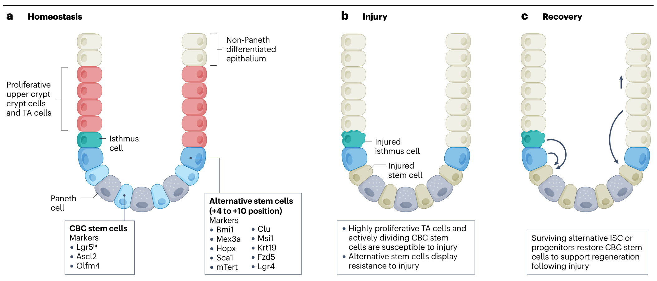
图 1 · 稳态与损伤再生。原文 PDF 第 3 页。
从类器官建立到生态位解析
肠干细胞研究的进展将标记鉴定、体外类器官培养、组织移植与空间生态位研究逐渐连接起来(PDF 第 4 页)。
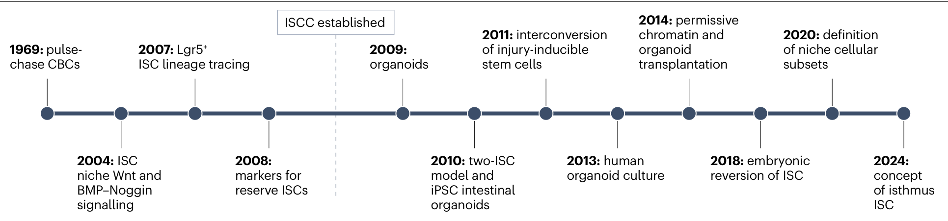
图 2 · 肠干细胞研究里程碑。原文 PDF 第 4 页。
空间生态位如何组织再生
trophocyte 提供 RSPO 与 BMP 抑制信号,telocyte 富含 BMP,空间分布帮助组织更新与分化的平衡。小肠与结肠的细胞构成、WNT 来源和组织结构也存在差异(PDF 第 3–5、7 页)。
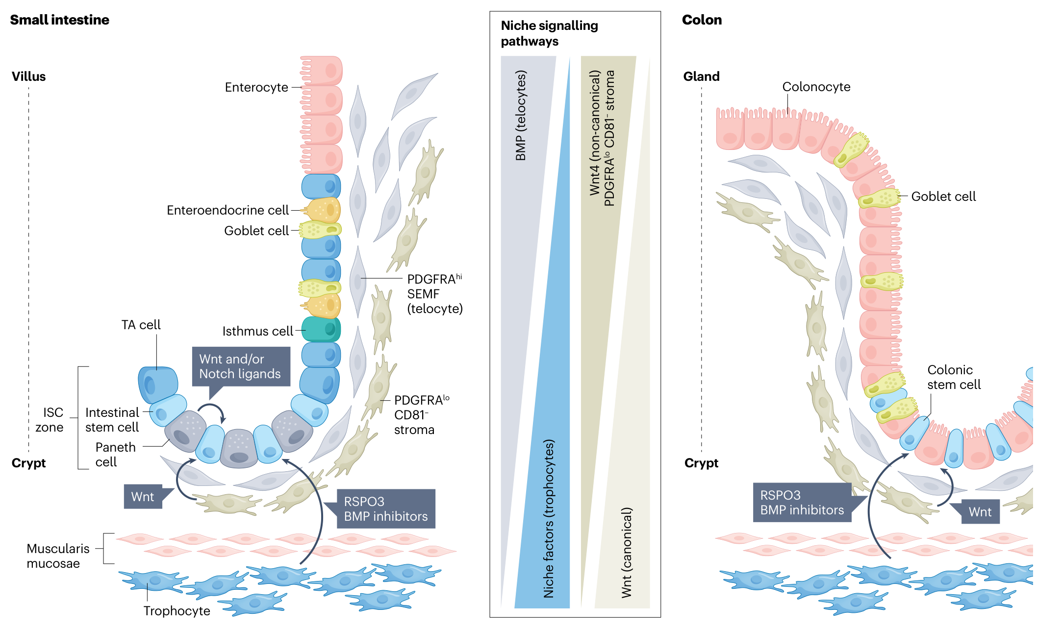
图 3 · 肠干细胞生态位的细胞组成。原文 PDF 第 7 页。
两类转化路径
| 路径 | 主要方案 | 正在解决的问题 |
|---|---|---|
| 补充细胞或组织 | 成人上皮类器官、iPSC 来源肠组织、含间质的复杂组织 | 植入、成熟度、供血与长期功能 |
| 调控再生环境 | EGF、WNT/RSPO、IL-22 等信号及其递送 | 局部作用、组合剂量与异常增殖风险 |
细胞产品的定义、规模化制造、免疫匹配与长期安全性,是从动物模型推进到人类应用的共同环节(PDF 第 8–10 页,表 4)。
值得持续关注的方向
- 人类疾病模型: 在模型中纳入间质和免疫组分,研究长期损伤与修复(第 5–8 页)。
- 空间信号来源: 将具体细胞亚群、分泌配体与上皮响应对应起来(第 3–7 页)。
- 组织工程: 根据疾病需求确定必要的支持细胞、支架与成熟度(第 9–10 页)。
原始文献线索:McCarthy 等关于肠道 BMP 信号梯度的研究(ref. 61);Sato 等单 Lgr5 干细胞类器官研究(ref. 44);Sugimoto 等类器官治疗短肠综合征的研究(ref. 48)。
主图与表格 · 7 张
Fig. 1 · PDF 第 3 页
Fig. 1 | Models of epithelial regeneration during homeostasis and injury repair in the small intestine. a, Proliferating Lgr5+ intestinal stem cells (ISCs) and slow-cycling putative ‘reserve’ ISCs occupy distinct locations within the crypt. Isthmus cells marked by Fgfbp1 and Lgr4 expression were identified as displaying potential ISC properties. b, In the context of various modes of injury, Lgr5+ crypt base columnar cells (CBCs) and many cells in the upper crypt are lost. c, Following such injury, regeneration occurs via survival of upper crypt cells or alternative stem cells and/or cellular plasticity of differentiated cells that reconstitute the epithelium and restore homeostasis. Light blue, CBC stem cells; dark blue, alternative stem cells; red, proliferative upper crypt cells and transit amplifying (TA) cells; grey, Paneth cells; beige, non-Paneth differentiated epithelium.
Fig. 2 · PDF 第 4 页
Fig. 2 | Timeline of ISC research. An overview of historical milestones in intestinal stem cell (ISC) research. The timeline highlights major discoveries that shaped current understanding of ISCs, beginning with pulse-chase studies of crypt base columnar cells (CBCs) in 1969 (ref. 13) and the identification of niche-associated Wnt and bone morphogenetic protein (BMP)–Noggin signalling in 2003–2004 (refs. 54–57). The field was further advanced by Lgr5+ ISC identification and lineage tracing in 2007 (ref. 14), the discovery of Bmi1 and other reserve ISC markers in 2008 (ref. 22) and the establishment of intestinal organoid cultures in 2009 (refs. 44,45). The two-ISC model with coexisting active and quiescent stem cells was proposed in 2010 (ref. 20), followed by functional demonstrations of injury-inducible stem cell interconversion16,17,28,112. Successful differentiation of human embryonic stem cells into intestinal organoids was obtained in 2011 (ref. 46). Additional landmarks include the establishment of human intestinal organoid culture43, regenerative plasticity by permissive chromatin42 and intestinal organoid transplantation47, post-injury fetal-like reversion of crypt stem cells38 and identification of niche cellular subsets61. The concept of isthmus–Fgfbp1+ ISCs18,19 expands the ISC hierarchy beyond classic models. iPSC, induced pluripotent stem cell; ISCC, Intestinal Stem Cell Consortium; LRCs, label-retaining cells.
Fig. 3 · PDF 第 7 页
Fig. 3 | Distinct cellular components in the ISC niches of small intestine and colon. Diverse cell types surround the crypt epithelium in both small intestine and colon. Telocytes (PDGFRAhi subepithelial myofibroblasts; SEMFs) that are enriched at the crypt–villus junction extend long cytoplasmic processes and express abundant bone morphogenetic protein (BMP) transcripts. The PDGFRAlo mesenchyme is subdivided into CD81−and CD81+ subsets, with the latter representing trophocytes that reside below the muscularis mucosae and elaborate high levels of intestinal stem cell (ISC)-stimulatory signals such as RSPO3 and BMP inhibitors. The opposing signal gradients that originate in trophocytes and telocytes control ISC proliferation, differentiation and homeostasis. TA, transit amplifying.
Table 1 · PDF 第 5 页
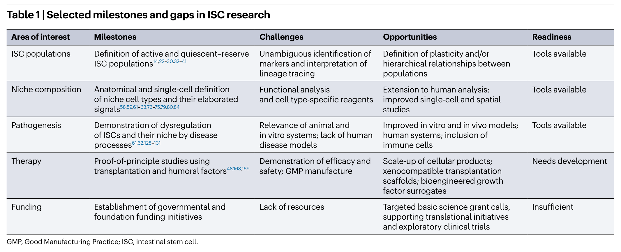
表题与脚注见截图。
Table 2 · PDF 第 6 页
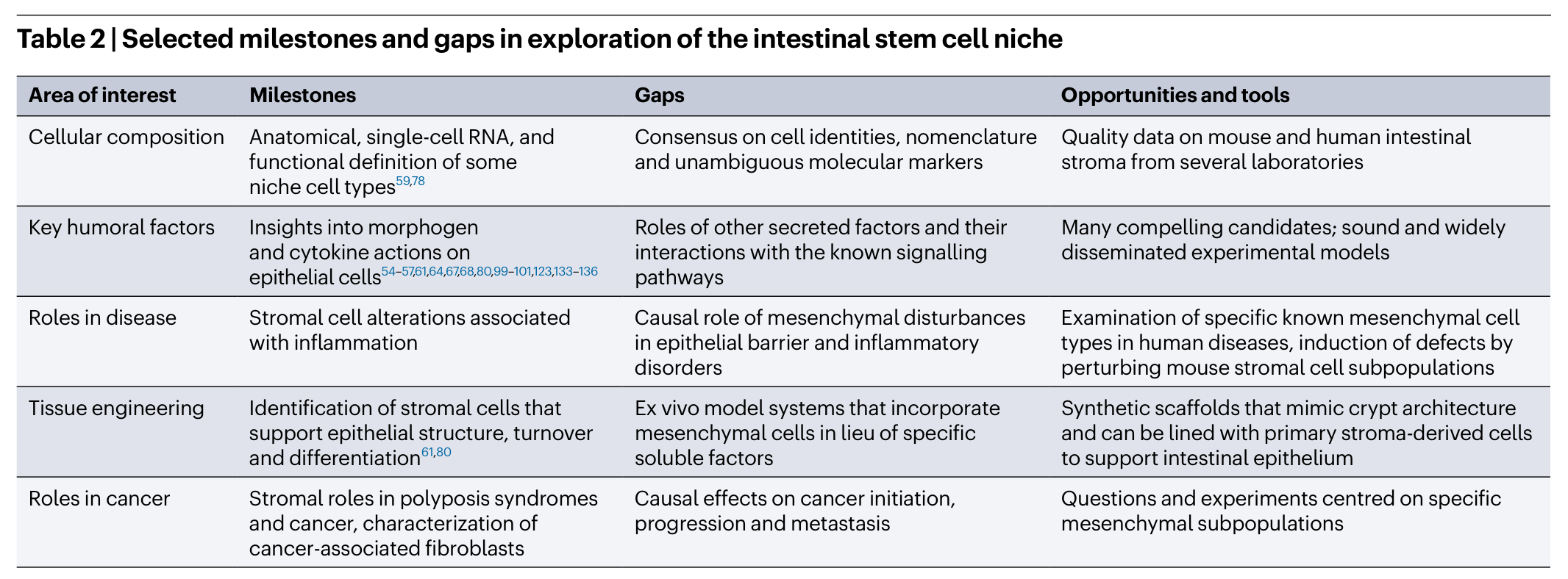
表题与脚注见截图。
Table 3 · PDF 第 8 页
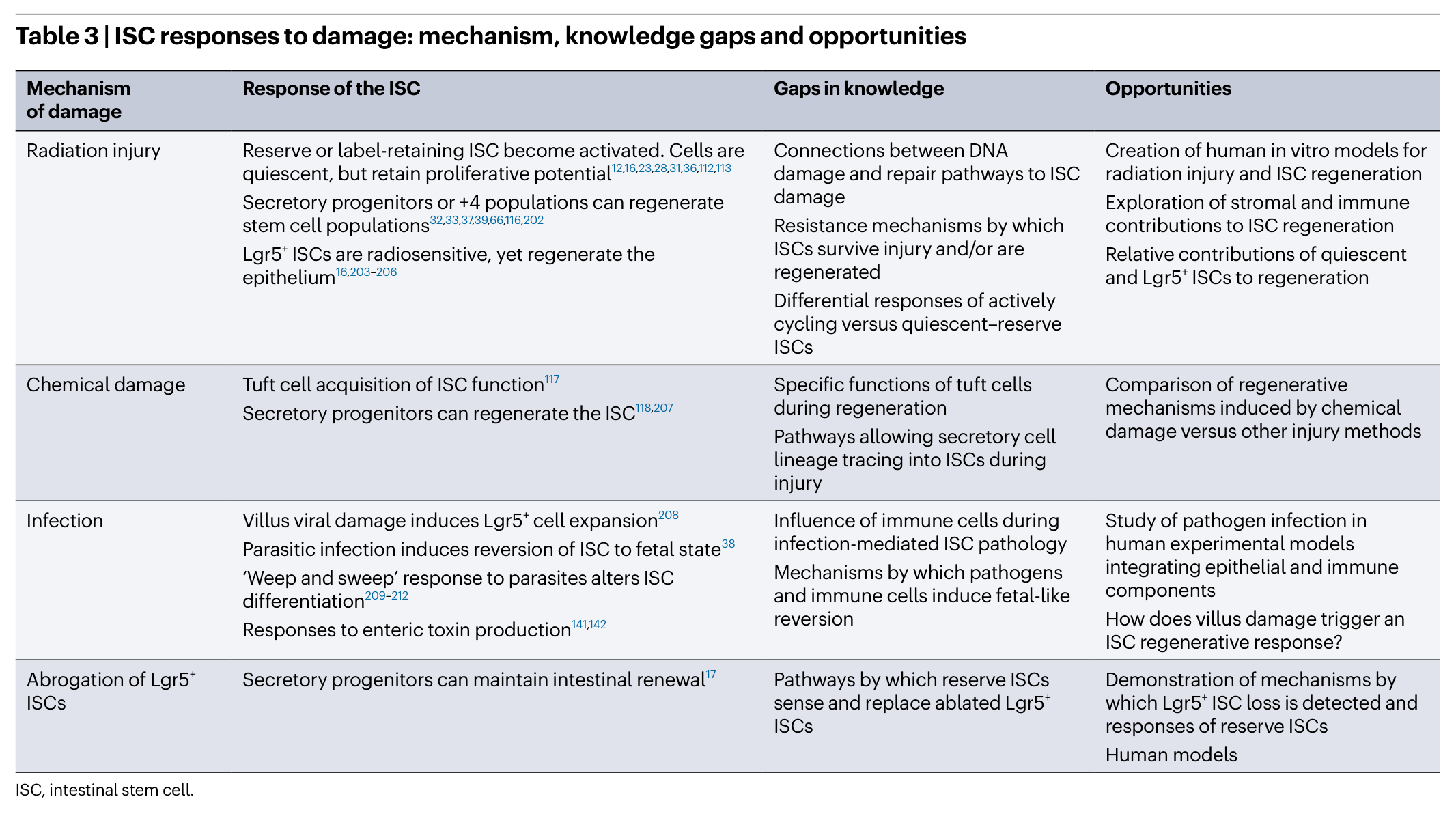
表题与脚注见截图。
Table 4 · PDF 第 10 页
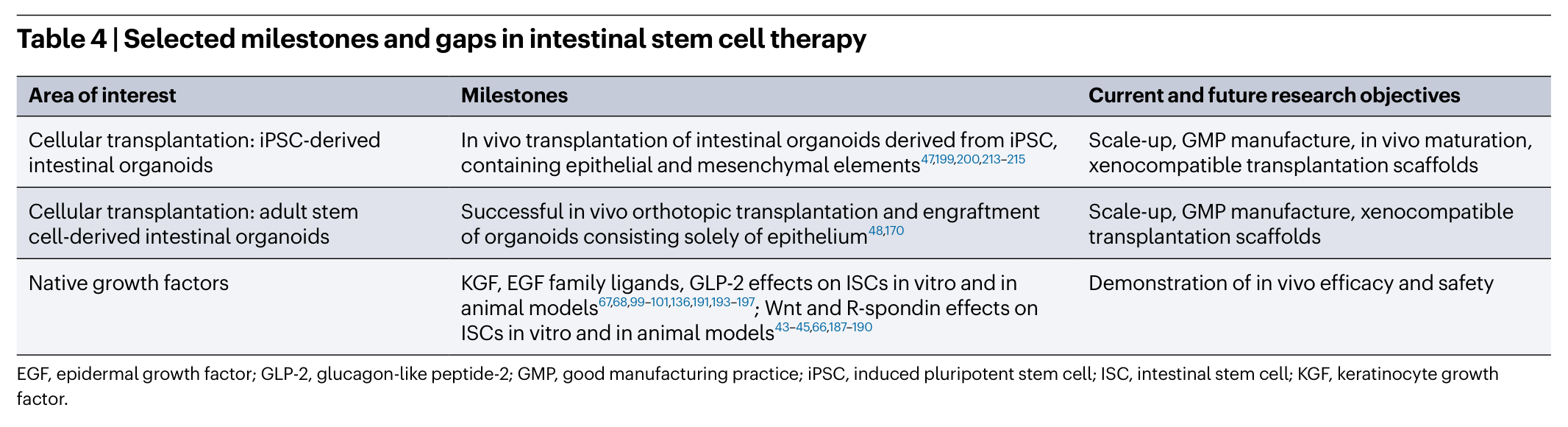
表题与脚注见截图。
完整阅读记录与证据表
文献卡:Building and regenerating intestines by manipulating intestinal stem cells
- Paper ID:p-0004-rev-building-regenerating-intestines
- 分析日期:2026-09-08;技能:review-read
- 原文类型:Roadmap;期刊:Nature Reviews Gastroenterology & Hepatology
- 阅读覆盖:PDF 共14页。第1–10页正文及第11页结论末段已读;表1–4及图1–3图注已读;图1(第3页)与图3(第7页)已视觉查看,图2时间轴仅正文/图注。第11–14页参考文献仅定位追读条目,第14页利益冲突已读。所引原始论文尚未直接核查。
- 人工核对:pending(待核对)
- 本卡页码均为从1开始的PDF页序。
一句话结论
该Roadmap把肠道再生的重点从单一干细胞标记扩展到“细胞状态—空间生态位—组织重建”,并提出细胞移植和生态位信号调控两类转化路径;大多数候选方案仍须跨越人类有效性、安全性和制造验证(PDF第2–10页,图1、3与表1–4)。
研究问题与背景
如何把肠干细胞(ISC)的更新与损伤修复能力变成可用治疗?作者强调ISC状态受到局部生态位调节,疾病既可能损伤上皮,也可能破坏维持再生的间质、免疫、血管和神经支持(第2–8页)。
贡献与比较对象
文章由ISCC成员总结联盟及领域进展,按“进展—挑战—机会”组织。对比活跃Lgr5+ CBC、候选储备细胞/峡部祖细胞、损伤可塑性模型,以及成人来源类器官、iPSC来源组织和包含间质的复杂培养系统(第2–5、8–10页)。这不是同一实验中的优劣比较。
范围与选文方法
原文类型是Roadmap,本地按rev流程归档为叙述性路线图。ISCC资助周期2009–2024年是合作项目时段,不是本综述检索截止日期(第1、3页)。未报告系统检索式、正式纳排标准或Meta分析,不能冠以系统综述。本文引用的原始研究均为转述。
分类体系与代表工作
| 主张或结果 | 数字、单位、条件 | PDF页序/图表 | 依据类型 | 核对状态 |
|---|---|---|---|---|
| ISC具有自我更新与多向分化能力 | 作者概括人肠上皮约5–7天更新 | 第2页 | 转述 | 待人工核对;不是本文新测定 |
| 稳态与损伤再生细胞来源有争议 | Lgr5+ CBC、候选储备细胞、Fgfbp1+峡部细胞及分化细胞可塑性 | 第2–3页图1 | 转述+作者模型;图已查看 | 标记特异性需核对 |
| 空间生态位决定更新/分化平衡 | trophocyte提供RSPO及BMP抑制信号;telocyte富含BMP,不能把全部间质视为同一种细胞 | 第3–5、7页图3 | 转述+概念图已查看 | 待人工核对 |
| 小肠与结肠不可互换 | Paneth细胞及WNT来源、杯状细胞比例和组织结构存在差异 | 第4–5、7页图3 | 转述 | 待人工核对 |
| 损伤机制影响再生来源 | 放射、化学、感染、直接消融Lgr5+细胞应分开讨论 | 第6–8页表3 | 转述/表格文字 | 不能跨模型直接推广 |
| 细胞治疗包括不同材料体系 | 成人上皮类器官、iPSC肠组织、含间质的复杂组织、工程化支架 | 第8–10页表4 | 转述 | 动物植入不等于临床疗效 |
| 体液/药理策略尝试模拟生态位 | EGF、WNT/RSPO、IL-22等;局部/全身递送及组合剂量仍有问题 | 第9–10页 | 转述+作者建议 | 未独立核查药物状态 |
| 临床转化需要定义最小产品 | GMP、无异种成分、规模化、免疫匹配、长期植入和安全性 | 第9–10页表4 | 作者路线图 | 待验证 |
争议与开放问题
- 分化细胞“去分化”的谱系证据是否来自成熟细胞?作者指出Alpi、Fabp2、Krt20、Dll1等标记也可在部分峡部祖细胞中表达,CreER标记并不自动证明起点是终末分化细胞(第2页)。
- 简化模型能否复现人类损伤修复?小鼠与人、小肠与结肠、稳态与损伤不能互相替代;细胞亚群的命名和标记尚需共识(第4–7页,表2、Box1)。
- 应直接补入上皮,还是移植完整组织?最小必要支持细胞、组织成熟度、支架和供血/神经/免疫整合需按具体疾病定义(第9–10页)。
- 促进再生与避免异常增殖如何兼顾?局部限制与持续生长因子刺激的风险仍需研究(第9–10页)。
局限
作者明确指出
人类疾病模型不足;Cre表达范围不精确;非间质生态位组分研究不足;长期损伤再生及空间配体来源尚不清楚;候选疗法与已证实临床疗效之间仍有明显距离(第5–10页)。
AI 推断与理由
这份路线图适合建立问题清单,但不适合用来估计任何具体方案的疗效。第9页引述其他器官多能干细胞临床经验,作者同时指出所引综述中没有胃肠疾病应用,不能据此宣称肠道iPSC治疗已有普遍安全保证。第14页披露部分作者具有相关咨询或专利利益,应与临床证据等级一起阅读。
优先追读清单
未给定课题,按基础定义、生态位因果证据与转化代表性排序。全部为尚未核查的原始论文,标题取自本篇参考文献。
- Barker et al. Identification of stem cells in small intestine and colon by marker gene Lgr5(2007;第11页ref.14):明确稳态干细胞证据起点。
- McCarthy et al. Distinct mesenchymal cell populations generate the essential intestinal BMP signaling gradient(2020;第11页ref.61):核对空间生态位模型。
- Sato et al. Single Lgr5 stem cells build crypt–villus structures in vitro without a mesenchymal niche(2009;第11页ref.44):区分外源因子替代与体内生态位需求。
- Spence et al. Directed differentiation of human pluripotent stem cells into intestinal tissue in vitro(2011;第11页ref.46):理解iPSC/多能干细胞路线与成人类器官的差异。
- Sugimoto et al. An organoid-based organ-repurposing approach to treat short bowel syndrome(2021;第11页ref.48):核对功能改善、植入方式及适用边界。
待核对问题与后续阅读
补核图2时间轴视觉;核查谱系追踪标记的特异性;对拟采用方案回到原始论文看功能终点。在线发表日期仍为占位符;年份来自PDF版权2026,不把ISCC周期当发表日期。
人工补充
待人工填写;本次未创建或覆盖 discussion.md。