研究概述
脂肪组织可以提供营养、激素和炎症信号,但它也可能主动限制肿瘤。Curtin等人的研究提出:非肥胖状态下的乳腺脂肪细胞会分泌具有抑癌作用的氧脂素;肥胖时,这一保护信号减弱。(PDF第1–3页)
研究将活性指向亚油酸衍生物 9S-HODE(9S-羟基十八碳二烯酸):分泌物和组织中测到的是不区分手性的9-HODE,而外加9S标准品可引发乳腺癌细胞死亡及脂质过氧化,支持铁死亡机制。脂滴形成的保护作用主要在小鼠乳腺上皮细胞NMuMG和乳腺癌细胞E0771中得到功能验证。小鼠局部给药与患者来源类器官进一步显示抑癌潜力,证据仍处于临床前阶段。(图3–5)
脂肪细胞分泌物与肿瘤生长
高脂饮食诱导肥胖后,E0771、Py230和MCF7乳腺肿瘤模型的生长曲线均高于非肥胖对照。作者进一步培养分离出的成熟脂肪细胞,将其条件培养基与新鲜培养基按 1∶1 混合,观察肿瘤细胞的反应。
关键比较是普通培养基、非肥胖脂肪细胞分泌物和肥胖脂肪细胞分泌物三组:非肥胖乳腺脂肪细胞的分泌物降低E0771生长,而肥胖组接近普通培养基。这为“非肥胖组主动抑制”提供了依据。腹股沟脂肪细胞的分泌物在7种小鼠或人乳腺癌细胞系中呈现类似作用;性腺周围脂肪的效果不同,提示脂肪库的位置同样重要。(图1G–H)
把非肥胖小鼠的腹股沟脂肪细胞与MCF7共同接种到受体小鼠侧腹,也能减慢肿瘤生长;肥胖脂肪细胞没有同样作用。这是侧腹共接种实验,与前面的乳腺原位模型应分开理解。(图1I)
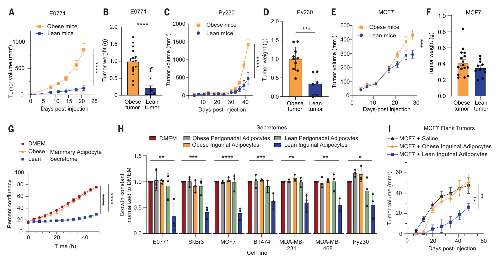
图1|原位肿瘤、不同脂肪库的条件培养基,以及脂肪细胞共接种实验。原文PDF第3页。
图1H的n=3代表独立脂肪细胞分离制备,相关筛选涵盖7种细胞系。后续大量分泌组实验采用雄鼠腹股沟脂肪,关键发现再用雌鼠乳腺脂肪验证。
铁死亡的功能证据
转录组首先给出线索:非肥胖分泌物处理后的E0771细胞富集铁死亡相关基因,NES=1.79,校正P=0.016。真正支撑机制的是几种互补读出和功能救援。(图2A–J)
| 证据 | 观察 | 说明 |
|---|---|---|
| 细胞死亡染料 | 多种乳腺癌细胞的Cytotox或SYTOX阳性增加 | 生长下降伴随细胞死亡 |
| 药物救援 | Ferrostatin-1或liproxstatin-1缓解生长抑制;所测凋亡、坏死性凋亡抑制剂未能救援 | 支持铁死亡参与生长抑制 |
| 脂质过氧化 | C11-BODIPY氧化信号增加,非肥胖小鼠肿瘤4-HNE水平较高 | 细胞及肿瘤中存在脂质氧化压力 |
| 铁稳态 | CD71/TFRC、HMOX1上调,RhoNox-1检测的Fe²⁺信号增加 | 铁代谢变化与死亡表型相伴 |
在体内,从肿瘤接种当天开始每日给予liproxstatin-1,非肥胖小鼠的E0771肿瘤长得更快,肥胖组未见同样的显著变化。这支持内源性铁死亡压力参与限制非肥胖环境中的肿瘤生长;干预与肿瘤接种同步启动。(图2L–N;方法PDF第10–11页)
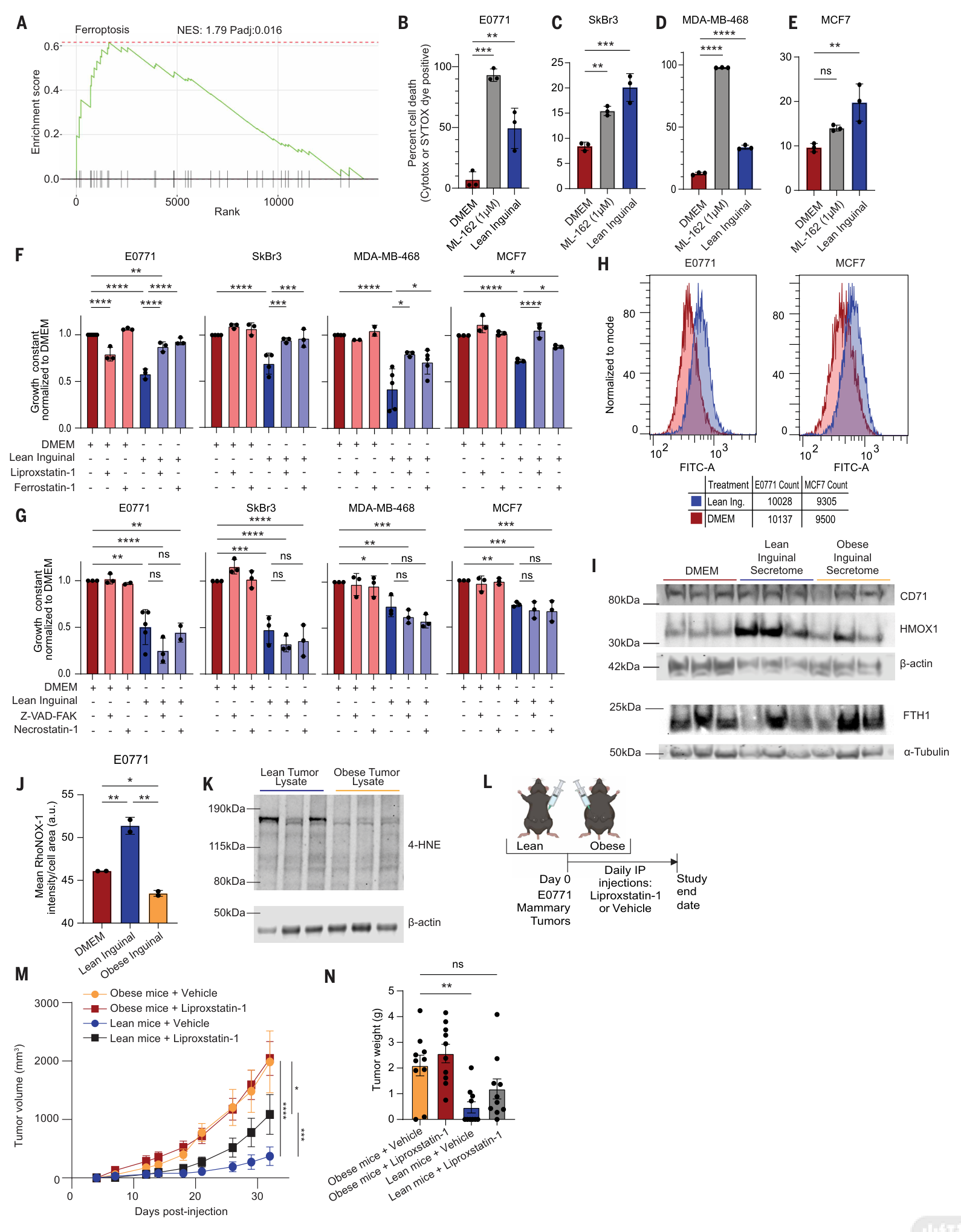
图2|细胞死亡、脂质过氧化、铁稳态及体内铁死亡抑制实验。原文PDF第4页;完整图注在第5页。
9S-HODE的筛选与验证
加热没有破坏分泌物的抑癌作用;活性却会被某些去脂处理消除。不同去脂材料的效果不同,作者据此把功能筛选与脂质组学交叉:先选出10个候选脂质特征,再用靶向质谱和外加化合物实验缩小范围。(图3A–F)
9S-HODE能够抑制E0771生长,而9R-HODE和13S-HODE在同一筛选条件下没有同样效果。同样的碳链,仅羟基位置或立体构型不同,活性就发生变化。外加9S-HODE还提高细胞死亡、脂质过氧化和Fe²⁺信号。针对该化合物的FSP1过表达救援由主文引用补图S7I报告;目前尚未取得该补图,FSP1救援按正文转述保留。(图3F–L;PDF第5页)
质谱定量不区分手性,组织和分泌物中测得的是9-HODE;9S构型的功能特异性来自外加标准品比较。(PDF第5页)
检测浓度与外加实验剂量:图3D中非肥胖分泌物的9-HODE约为300 pmol/mL,即约0.3 μM(读图估计),用于生长实验时还与新鲜培养基按1∶1混合;图3G–I的外加9S-HODE为20或60 μM。细胞有效暴露还受样品、手性和脂质结合状态影响。作者报告“按非肥胖分泌物浓度补回即可恢复抑制”,但证据在尚未取得的补图S9A,本轮尚未独立核读。(PDF第7、11页)
在脂肪细胞中抑制LOX,尤其使用5-LOX抑制剂zileuton,会降低9-HODE分泌并减弱条件培养基的生长抑制作用。非肥胖荷瘤小鼠口服zileuton后,E0771生长也加快。这是对5-LOX依赖性的药理支持,体内实验采用全身给药。(图3M–O)
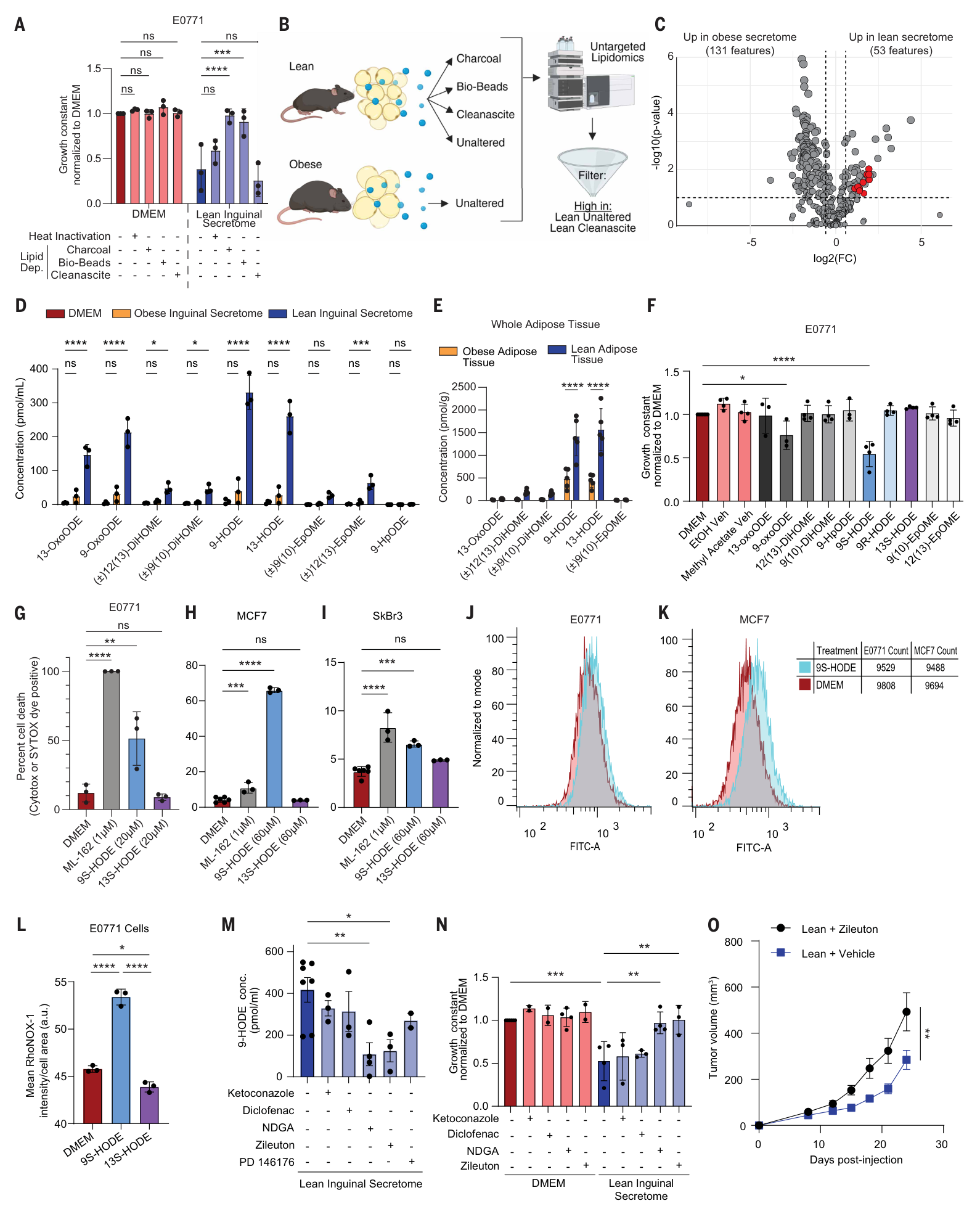
图3|去脂筛选、氧脂素定量、立体构型比较与5-LOX药理干预。原文PDF第6页;图注跨第6–7页。
脂滴的保护作用
在非肥胖脂肪细胞分泌物处理下,正常小鼠乳腺上皮细胞NMuMG和EpH4的总体生长受影响较小。NMuMG出现短暂生长停顿,随后恢复,并伴随脂滴增加。作者进一步从两个方向检验脂滴的保护作用:(图4C–L)
- 抑制NMuMG细胞中的DGAT1/2,脂滴积累减少,细胞对非肥胖脂肪细胞分泌物的生长抑制变得敏感。
- 在E0771中增加DGAT2表达,脂滴形成增多,分泌物造成的生长抑制得到救援。
这把选择性与脂质储存能力联系起来:脂滴可能将有害脂质隔离,降低其引发膜脂过氧化的机会。DGAT2救援也提示,肿瘤自身的脂滴代谢可能影响对这类干预的敏感性。
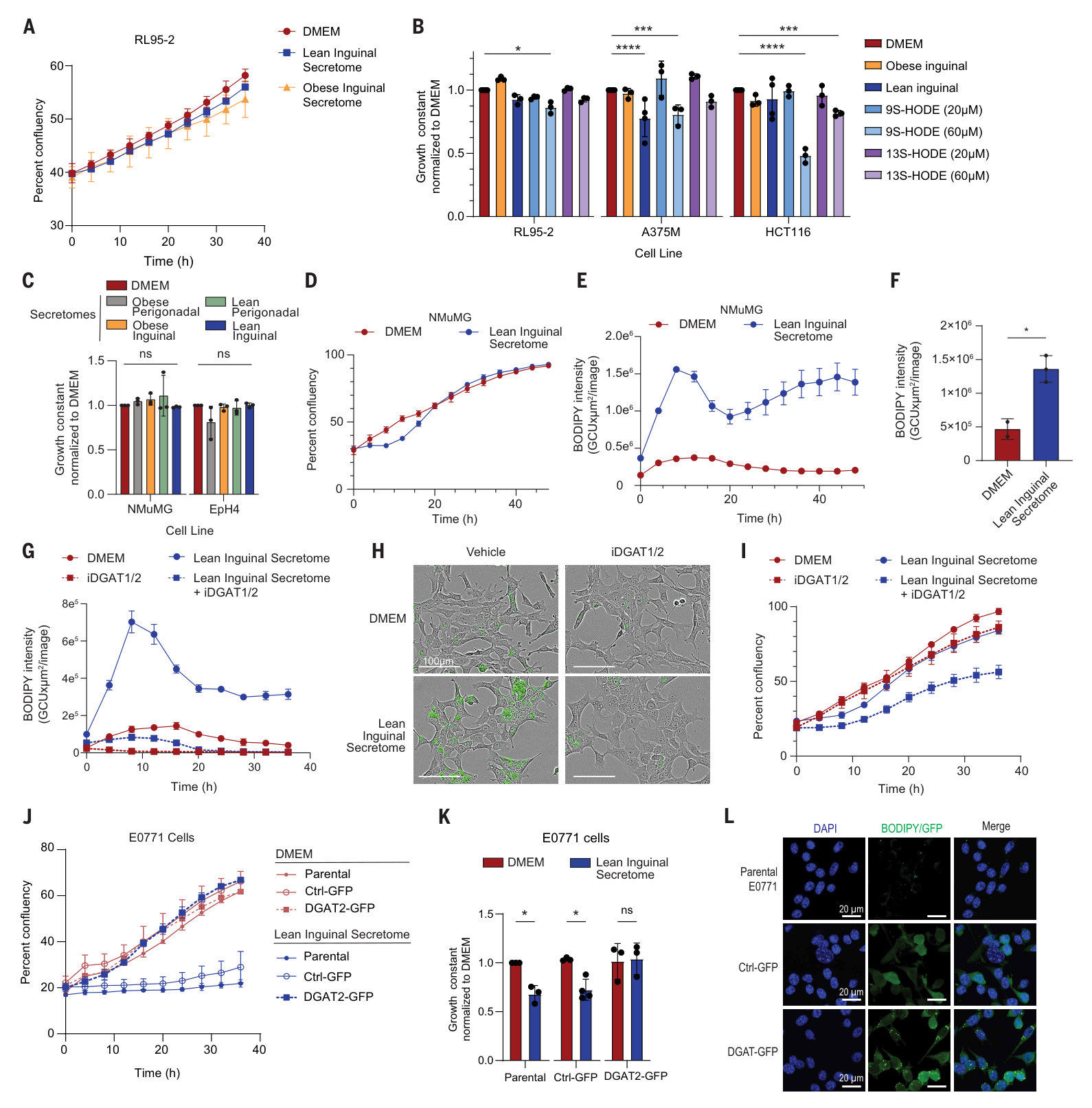
图4|不同细胞的敏感性,以及DGAT抑制和DGAT2过表达实验。原文PDF第8页。
模型间的敏感性存在差异。图4B中,HCT116结直肠癌细胞在60 μM 9S-HODE下也有明显生长下降,其他细胞在部分条件下也有变化。不同细胞模型、浓度和脂质处理能力共同影响敏感性。
小鼠局部给药与人源材料
作者在肿瘤达到 80–150 mm³ 后随机分组,对肥胖小鼠每周进行3次瘤内注射。方法写明每次注射液为 100 μM、100 μL 的9S-HODE或匹配溶剂;这是局部给药浓度和体积。E0771、MDA-MB-468和MCF7模型呈现生长抑制;Py230的用药组平均生长曲线较低,但图5C–D未给出相关比较的显著性标记或确切P值。(图5A–H;PDF第11页)
不同终点的证据强度也有差别:E0771终点瘤重的肥胖用药组与肥胖溶剂组比较P=0.0632,尽管生长曲线显示差异;Py230终点瘤重未标显著性。各模型以肿瘤生长和重量为主要读出。
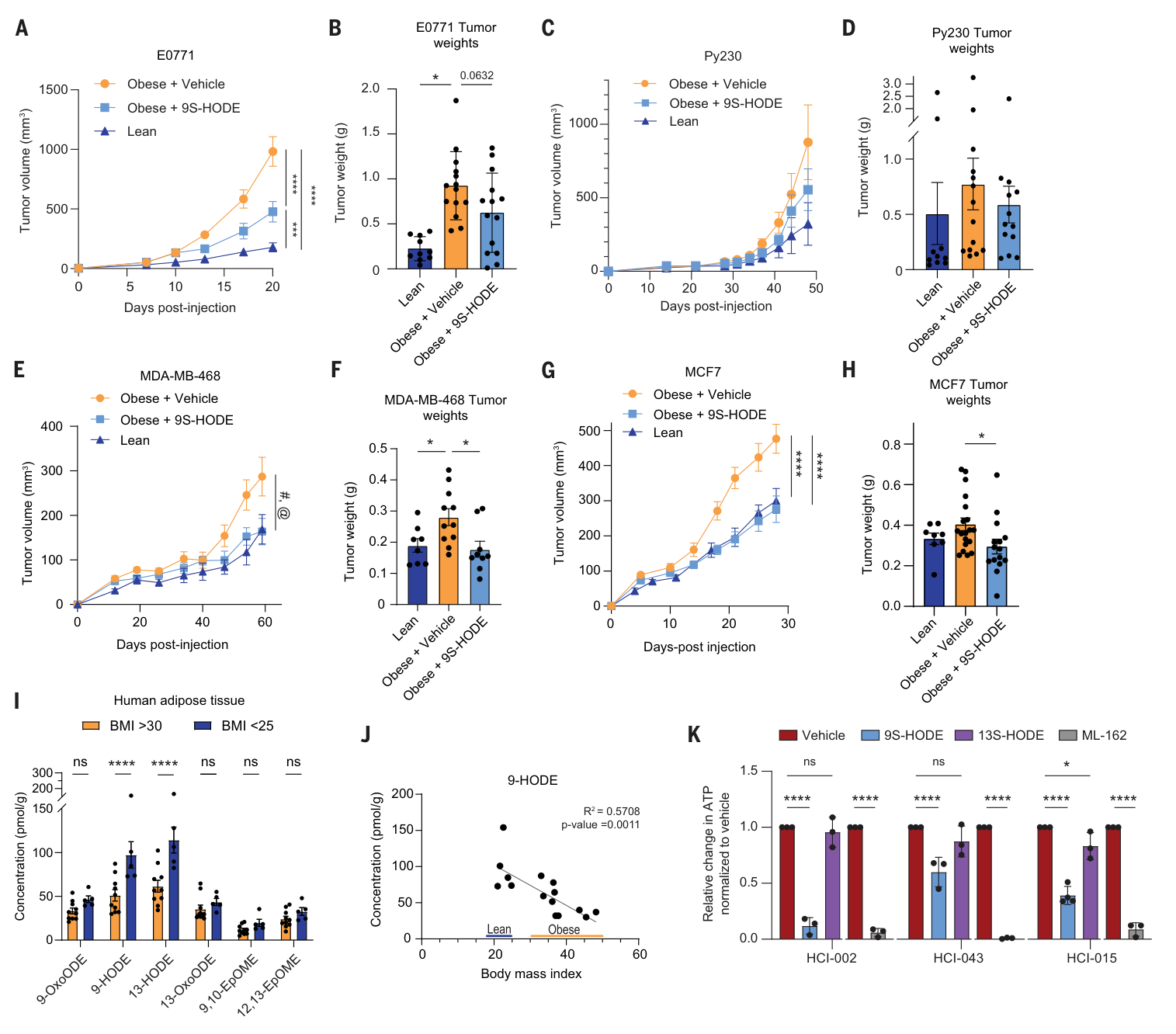
图5|四种肿瘤模型的局部给药、人乳腺脂肪中的9-HODE及患者来源类器官。原文PDF第9页。
人源证据分为两部分:
| 材料 | 主要发现 | 证据层级 |
|---|---|---|
| 乳腺脂肪组织 | 9-HODE与供体BMI负相关,R²=0.5708,P=0.0011;每个点代表同一供体左右乳腺测量的平均 | 人体组织关联,未检验癌症风险或患者治疗结局 |
| HCI-002、HCI-043、HCI-015三种模型 | 9S-HODE降低CellTiter-Glo 3D的ATP读数 | 来自三阴性乳腺癌患者来源异种移植瘤的类器官,属于离体药物反应 |
三种类器官对9S-HODE的反应幅度不同。13S-HODE总体作用弱得多,但图5K中HCI-015仍标有轻度显著变化。这些类器官实验主要报告ATP读数变化。(图5I–K;方法PDF第10页)
局限与未解问题
这项研究提出并以多组实验支持一条脂肪组织保护机制:非肥胖脂肪细胞的氧脂素信号 → 肿瘤细胞铁与脂质稳态变化 → 铁死亡压力 → 生长受限;DGAT依赖的脂滴形成可以缓冲这一过程。局部给药实验提供了进一步开发的线索。信号的直接受体、肥胖为何降低其产生、系统给药的暴露与安全性,以及哪些患者最可能敏感,仍有待解决。
机制上,9S-HODE直接受体以及CD71、HMOX1的必要性尚待验证;作者提出的立体构型识别受体属于假说。全身5-LOX抑制的来源归属也需脂肪细胞特异干预进一步明确(PDF第9–10页)。
转化方面,分泌物检测与外加标准品存在浓度、手性及结合状态差异,生理浓度补回证据仍待补图核查;人源类器官主要使用ATP读出,铁死亡机制和人体安全性需要后续验证。局部给药、早期机制干预与系统治疗分别具有不同的暴露与观察条件(图2–5;PDF第10–11页)。
本文导读依据桌面正式版PDF全部18页与5张主图。正式版补充图S1–S9、MDAR清单及Data S1–S7尚未取得,主文引用这些材料的细节未独立核读;相关缺项保存在阅读记录中。
主图与表格 · 5 张
fig-1.png · PDF 第 3 页
Fig. 1. Lean mammary adipocytes inhibit breast cancer cell growth. (A to F) Orthotopic tumor growth is accelerated in obese mice. (A) Growth kinetics of E0771 mammary tumors in female C57BL/6J mice fed a high-fat (obese; n = 16) or standard chow (lean; n = 16) diet. (B) End-point tumor burden. (C) Growth kinetics of Py230 mammary tumors in female C57BL/6J mice fed a high-fat (obese; n = 10) or standard chow (lean; n = 6) diet. (D) End-point tumor burden. (E) Growth kinetics of MCF7 mammary tumors in NRG mice fed a high-fat (obese; n = 16) or standard chow (lean; n = 11) diet. (F) End-point tumor burden. (G) Representative growth curves demonstrating that the secretomes of lean, but not obese, mammary adipocytes suppress breast cancer cell growth. (H) Secretome from lean inguinal adipocytes decreases the growth constant across seven breast cancer cell lines. Secretomes from obese inguinal or perigonadal (lean or obese) adipocytes do not inhibit breast cancer cell growth (n = 3 independent adipocyte isolations). (I) Coinjection of lean inguinal adipocytes with MCF7 breast cancer cells reduces tumor growth compared with coinjection with obese adipocytes or a saline control (n = 6). [(A) to (F) and (I)] Data are means ± SEMs. [(G) and (H)] Data are means ± SDs. P values calculated using two-way ANOVA followed by Šídák’s multiple comparisons test [(A), (C), and (E)] or followed by Tukey’s multiple comparison test [(G) and (I)], calculated using unpaired t test [(B), (D), and (F)], or calculated using one-way ANOVA followed by Dunnett’s multiple comparisons test (H). *P < 0.05; **P < 0.01; ***P < 0.001; ****P < 0.0001.
fig-2.png · PDF 第 4 页
Fig. 2. Lean adipocytes induce ferroptosis in breast cancer cells. (A) Gene set enrichment analysis for ferroptosis-related genes in E0771 cells treated with the lean inguinal adipocyte secretome compared with DMEM control. (B to E) Lean inguinal secretome and the ferroptosis inducer ML-162 increase cell death in E0771 cells (B), SkBr3 cells (C), MDA-MB-468 cells (D), and MCF7 cells (E), measured by Cytotox or SYTOX dye incorporation (n = 3). (F and G) Pharmacological inhibition of ferroptosis (F), but not other cell death pathways (G), rescues the growth of breast cancer cells treated with the lean inguinal secretome (n = 3 to 5). (H) Representative C11-BODIPY staining in E0771 and MCF7 breast cancer cells shows increased lipid peroxidation after treatment with lean inguinal secretome. (I) E0771 cells up-regulate iron-related proteins CD71/TFRC and HMOX1 in response to lean versus obese inguinal secretome (n = 3). (J) Intracellular Fe2+ levels are increased in E0771 cells treated with lean inguinal secretome compared with DMEM control medium or obese inguinal secretome, quantified by mean cellular RhoNox-1 fluorescence intensity (n = 2). (K) Immunob- lot analysis of 4-HNE in E0771 tumor lysates shows increased 4-HNE abundance in tumors from lean versus obese mice (n = 3 mice). (L to N) (L) Schematic of experimental design. (M) Daily injection of the ferroptosis inhibitor liproxstatin-1 accelerates mammary E0771 tumor growth in lean mice (n = 8) compared with vehicle control (n = 6), with no effect observed in obese mice with (n = 10) or without (n = 10) liproxstatin-1. (N) End-point tumor weights. [(B) to (G) and (J)] Data are means ± SDs. [(M) and (N)] Data are means ± SEMs. P values calculated using one-way ANOVA followed by Dunnett’s multiple comparison test [(B) to (E)], Šídák’s multiple comparisons test [(F) and (G)], or Tukey’s multiple comparisons test [(J) and (N)] or calculated using two-way ANOVA followed by Šídák’s multiple comparisons test (M). ns, not significant; *P < 0.05; **P < 0.01; ***P < 0.001; ****P < 0.0001.
fig-3.png · PDF 第 6 页
Fig. 3. Lean adipocytes secrete ferroptosis inducer 9S-HODE. (A) The growth-inhibitory effect of lean inguinal adipocyte secretome is not mediated by a heat-labile protein. Lipid depletion using charcoal dextran or Bio-Beads SM-2 Adsorbents, but not Cleanascite, restores E0771 growth (n = 3). (B) Schematic of lipidomics workflow. (C) Volcano plot of lipid features comparing lean and obese inguinal adipocyte secretomes (mean of three independent secretomes per group). Features enriched in lean versus obese secretome and depleted by charcoal dextran and Bio-Beads SM-2 Adsorbents, but not Cleanascite, are highlighted in red. (D and E) Quantification of candidate oxylipins in lean versus obese inguinal adipocyte secretome and DMEM control (n = 3 independent secretomes) (D) or lean versus obese female mammary white adipose tissue (n = 5) (E). (F) Treatment of E0771 cells with candidate lipids identifies 9S-HODE and 9-OxoODE as sufficient to inhibit E0771 growth (n = 4). (G to I) 9S-HODE and ML-162 induce cell death in E0771 cells (G), MCF7 cells (H), and SkBr3 cells (I) compared with DMEM or 13S-HODE controls (n = 3). (J and K) Representative C11-BODIPY staining in E0771 cells (J) and MCF7 cells (K) showed increased lipid peroxidation after 9S-HODE treatment (20 or 60 μM, respectively) compared with DMEM control. (L) Intracellular Fe2+ levels are increased in E0771 cells treated with 9S-HODE compared with 13S-HODE or DMEM control (n = 3). (M) Relative concentration of 9-HODE in the secretome of lean inguinal adipocytes was reduced after treatment of adipocytes with NDGA or zileuton (n = 3 to 7). (N) The growth-inhibitory effect of lean inguinal adipocyte secretome on E0771 cells is lost when adipocytes were treated with NDGA or zileuton. (O) Daily administration of zileuton accelerates E0771 mammary tumor growth in lean mice (n = 10) compared with vehicle control (n = 5). [(A), (D) to (I), (L), and (N)] Data are means ± SDs. [(M) and (O)] Data are means ± SEMs. P values calculated using two-way ANOVA followed by Dunnett’s multiple comparison test [(A) and (D)] or Šídák’s multiple comparisons test [(E) and (O)] or calculated using one-way ANOVA followed by Dunnett’s multiple comparison test [(F) to (I), (M), and (N)] or Tukey’s multiple comparisons test (L). *P < 0.05; **P < 0.01; ***P < 0.001; ****P < 0.0001.
fig-4.png · PDF 第 8 页
Fig. 4. Normal mammary epithelial cells are insensitive to lean adipocytes. (A and B) Representative growth kinetics of RL95-2 (A) and growth constant of RL95-2 (endome- trial), A375M (melanoma), and HCT116 (colorectal) cancer cell lines (B) show little to no sensitivity to lean inguinal adipocyte secretome or 9S-HODE at concentrations that strongly suppress breast cancer cell growth (n = 3). (C) Lean inguinal adipocyte secretome does not alter the growth constant of normal mammary epithelial cell lines NMuMG and EpH4 (n = 3). (D) Representative growth curve showing a transient growth arrest in NMuMG cells after treatment with lean inguinal adipocyte secretome. (E and F) Lean inguinal adipocyte secretome increases lipid droplet accumulation. (E) Representative time course of intracellular BODIPY fluorescence intensity. (F) Quantification of BODIPY fluorescence intensity 8 hours after treatment with lean inguinal adipocyte secretome or DMEM control (n = 2 to 3). (G to I) DGAT1/2-mediated lipid droplet accumulation is required for mammary epithelial cell protection from the lean inguinal adipocyte secretome. (G) Representative BODIPY fluorescence intensity over time with or without DGAT inhibition (iDGAT1/2). (H) Representa- tive images at 8 hours posttreatment. Scale bars, 100 μm. (I) Representative growth curves showing increased sensitivity of NMuMG cells to lean inguinal adipocyte secretome with DGAT1/2 inhibition. (J to L) E0771 cells stably expressing DGAT2 are resistant to the lean inguinal adipocyte secretome. (J) Representative growth curves. (K) Quantification of growth rate constants (n = 3 to 4). (L) Representative images showing increased lipid droplet accumulation labeled with BODIPY in DGAT2-GFP–expressing E0771 cells. Scale bars, 20 μm. DAPI, 4′,6-diamidino-2-phenylindole. [(A), (D), (E), (G), (I), and (J)] Data are means ± SDs of technical replicates. [(B), (C), (F), and (K)] Data are means ± SDs of biological replicates. P values calculated using two-way ANOVA followed by Dunnett’s multiple comparison test (B) or followed by Šídák’s multiple comparisons test (K) or calculated using one- way ANOVA followed by Dunnett’s multiple comparison test (C) or unpaired t test (F). *P < 0.05; **P < 0.01; ***P < 0.001; ****P < 0.0001.
fig-5.png · PDF 第 9 页
Fig. 5. 9S-HODE suppresses breast cancer growth. (A to H) Intratumoral administration of 9S-HODE suppresses breast cancer tumor growth. Orthotopic mammary tumors were treated three times per week with intratumoral injections of 9S-HODE or vehicle. Mice were randomized when tumors reached 80 to 150 mm3; lean mice were left untreated. E0771 mammary tumor growth (A) and end-point tumor weights (B) (n = 14 obese+vehicle, n = 14 obese+ 9S-HODE, n = 10 lean). Py230 mammary tumor growth (C) and end-point tumor weights (D) (n = 14 obese+vehicle, n = 13 obese+ 9S-HODE, n = 10 lean). MDA-MB-468 mammary tumor growth (E) (“#” indicates obese compared with lean, ****P < 0.0001; “@” indicates obese compared with obese 9S-HODE, ****P < 0.0001) and end-point tumor weights (F) (n = 11 obese+vehicle, n = 9 obese+ 9S-HODE, n = 8 lean). MCF7 mammary tumor growth (G) and end-point tumor weights (H) (n = 19 obese+vehicle, n = 15 obese+ 9S-HODE, n = 8 lean). (I and J) Human breast white adipose tissue from lean female donors (BMI < 25) have increased levels of 9-HODE compared with obese female donors (BMI > 30). (I) Abundance of select oxylipins in donor breast tissue, with left and right breast oxylipin levels averaged per donor. (J) 9-HODE concentrations as a function of donor BMI shows a significant inverse correlation between BMI and 9-HODE levels [coefficient of determination (R2) = 0.5708, P = 0.0011]; each data point is average of left and right breast 9-HODE levels per donor. (K) Breast cancer patient–derived xenograft organoids are sensitive to 9S-HODE treatment, quantified by Celltiter-Glo 3D for three TNBC patient–derived organoid lines. ATP, adenosine 5′-triphosphate. [(I) and (K)] Data are means ± SDs. [(A) to (H)] Data are means ± SEMs. P values calculated using one-way ANOVA followed by Dunnett’s multiple comparison test [(A), (C), (E), and (G)] or calculated using two-way ANOVA followed by Tukey’s multiple comparison test [(B), (D), (F), and (H)], followed by Šídák’s multiple comparisons test (I), or followed by Dunnett’s multiple comparisons test (K). *P < 0.05; **P < 0.01; ***P < 0.001; ****P < 0.0001.
完整阅读记录与证据表
文献卡:Lean adipocyte oxylipin signaling restrains breast cancer through ferroptosis
- Paper ID:p-0012-art-lean-adipocyte-oxylipin-signaling
- 分析日期:2026-09-16;技能:article-read、morning-report、pdf、web-publish(2026-09-16晨会)。
- 正式发表:Science 393(6816), eaea4287,2026-09-10;DOI:10.1126/science.aea4287。题名、9名作者、卷期、文章号由PDF第1、18页与PubMed 42721252核对。
- 读取范围:桌面主PDF全部18页。PDF第1页是研究摘要与图形摘要;第2–17页对应文章印刷页1–16;第18页为编辑摘要、出版信息与许可。主图1–5及完整图注均已阅读。
- 人工核对:pending。图集6个PNG(5主图、1图形摘要);完整原始图注见figures/captions.md,逐图状态见figures/README.md。
- 缺项:正式版Figs. S1–S9、MDAR Reproducibility Checklist、Data S1–S7未取得;GSE300839未下载重分析。补充材料阅读尚未完成;主文主体已分析,整体processing_status记partial。
一句话结论
本研究用条件培养基、脂质筛选、铁死亡救援与小鼠局部补充实验,支持非肥胖脂肪细胞的氧脂素信号参与限制乳腺癌生长;9S-HODE具有功能活性,肥胖时内源性9-HODE减少,但其手性组成、直接受体与临床治疗价值尚未确定。
研究问题与贡献
既往工作多寻找肥胖时增加的促癌因素。本文检验非肥胖脂肪细胞是否提供保护性分泌物,并连接脂肪库、分子构型、铁死亡和脂滴缓冲。普通培养基作为第三比较基线,使“非肥胖组主动抑制”区别于仅比较肥胖与非肥胖。体内机制抑制与成瘤后局部补充承担不同证据角色。(PDF第2–10页)
方法与验证
- 小鼠:C57BL/6J或NRG雌鼠建立乳腺原位肿瘤,肥胖由至少9周高脂饮食诱导。MCF7模型额外植入雌激素缓释片。乳腺接种为双侧第4脂肪垫,图注n按原报告记录,同鼠双侧病灶的统计层级仍待原始数据复核。(PDF第10页)
- 分泌物:成熟脂肪细胞培养1周后开始收集,每48小时取条件培养基;用于肿瘤生长实验时与新鲜培养基1∶1混合。多数机制实验取雄鼠腹股沟脂肪,雌鼠乳腺脂肪验证关键结果。(PDF第2、11页)
- 生长:Incucyte每4小时成像,共72小时;指数生长期拟合倍增时间,再计算ln(2)/倍增时间。主要读出为汇合度及拟合生长常数。(PDF第11页)
- 机制:Cytotox/SYTOX、C11-BODIPY、RhoNox-1、免疫印迹、铁死亡抑制剂、FSP1或DGAT2过表达等互补读出。FSP1主要证据在未取得的补充图中,主文陈述已读,原图未独立核读。(PDF第3–8、12–13页)
- 脂质鉴定:非靶向Q-TOF功能交叉筛选后使用靶向QQQ及标准品验证;初筛采用原始P<0.1,非多重校正的确认性检验。质谱不区分9-HODE手性。(PDF第5、13–16页)
- 干预区别:liproxstatin-1从接种日开始每日腹腔给药10 mg/kg;zileuton从接种前3日起每日灌胃100 mg/kg;9S-HODE在肿瘤80–150 mm³后随机分组,100 μM、100 μL瘤内注射,每周3次。(PDF第10–11页)
- 人源:乳腺脂肪来自缩乳手术或快速尸检项目;BMI分组为<25与>30,左右乳腺数据按供体平均。三种TNBC患者来源异种移植瘤类器官HCI-002/HCI-043/HCI-015,给药72小时后测CellTiter-Glo 3D。(PDF第7、9–10页)
核心证据
| 主张或结果 | 数字、单位、条件 | PDF页序/印刷页/图表 | 依据类型 | 核对状态 |
|---|---|---|---|---|
| 肥胖组肿瘤生长较快 | 图注n:E0771肥胖16/非肥胖16;Py230 10/6;MCF7 16/11;MCF7终点瘤重未标显著性 | PDF3/正文2/图1A–F | 图表、图注 | 图文已核;双侧肿瘤统计单位待核 |
| 非肥胖分泌物主动抑制 | 普通培养基基线;7种乳腺癌细胞系;n=3独立脂肪细胞分离制备;性腺周围脂肪不呈同样作用 | PDF2–3/图1G–H | 正文、图表 | 已核 |
| 脂肪细胞共接种抑制生长 | MCF7与雄鼠腹股沟脂肪细胞共接种,受体为雌性NRG侧腹;图注n=6 | PDF3、10/图1I | 正文、图表、方法 | 已核,区别于原位接种 |
| 铁死亡相关转录富集 | E0771非肥胖分泌物相对DMEM:NES 1.79,Padj 0.016 | PDF4/正文3/图2A | 图表 | 已核;RNA原数据未重算 |
| 多种死亡与救援证据 | 4种细胞死亡读出n=3;铁死亡药物救援n=3–5,凋亡/坏死性凋亡抑制剂未救援 | PDF4–5/图2B–G | 图表、图注 | 已核;药物名称存在原文拼写差异 |
| 铁和脂质氧化改变 | C11-BODIPY;CD71/HMOX1印迹n=3;E0771 RhoNox-1 n=2;瘤4-HNE n=3 | PDF4–5/图2H–K | 图表、图注 | 已核;未直接证明铁节点必需性 |
| 铁死亡限制非肥胖组体内生长 | liproxstatin-1:非肥胖用药8/溶剂6;肥胖用药10/溶剂10,按图注;从接种日起给药 | PDF4–5、11/图2L–N | 图表、方法 | 已核;不据“一个显著一个不显著”自行宣称正式交互检验 |
| 脂质筛选定位活性 | 10个候选特征;9S-HODE抑制,9R/13S同一筛选条件无同样作用;候选脂质功能试验n=4 | PDF5–7/图3A–F | 正文、图表 | 已核;内源性仅定量9-HODE |
| 外加9S-HODE复制死亡表型 | E0771死亡面板20 μM、MCF7/SkBr3 60 μM,n=3;C11图注E0771/MCF7 20/60 μM | PDF6–7/图3G–L | 图表、图注 | 已核;两种剂量分别对应所列细胞 |
| 5-LOX药理依赖 | NDGA/zileuton降低9-HODE并减弱分泌物活性;zileuton体内图注n=10对照n=5 | PDF5–7/图3M–O | 正文、图表 | 已核;无脂肪细胞特异遗传证据 |
| 不同细胞的剂量响应 | 图4B中HCT116在60 μM时生长常数约为DMEM的一半(目测量级,不作精确效应量);n=3 | PDF8/正文7/图4A–B | 原图 | 已核,正文“insensitive”概括需收窄 |
| 脂滴保护机制 | NMuMG/EpH4总体增长较保留;DGAT抑制降低脂滴并增加敏感性;E0771 DGAT2过表达救援n=3–4 | PDF7–8/图4C–L | 正文、图表 | 已核;正常细胞证据主要为小鼠细胞系 |
| 成瘤后瘤内补充可抑制生长 | E0771三组图注n=14/14/10;Py230 14/13/10;MDA468 11/9/8;MCF7 19/15/8(肥胖溶剂/肥胖9S/非肥胖) | PDF9/正文8/图5A–H | 图表、图注 | 已核;各终点统计支持不一 |
| E0771终点瘤重未达常用显著阈值 | 肥胖溶剂vs肥胖9S-HODE:P=0.0632;Py230瘤重图未标显著性 | PDF9/图5B、D | 原图 | 已核;没有把曲线差异等同所有终点显著 |
| 人乳腺脂肪相关性 | BMI<25、>30;9-HODE与BMI R²=0.5708,P=0.0011;每点为供体左右乳腺平均 | PDF7、9/图5I–J | 正文、图表 | 已核;未猜供体总n,数据S7未获 |
| 三种人源类器官反应 | 9S-HODE降低ATP,幅度不同;13S-HODE在HCI-015有轻度显著变化 | PDF9–10/图5K | 原图、方法 | 已核;无直接临床疗效/类器官铁死亡救援读出 |
局限
作者明确指出或方法直接披露
- 9-HODE质谱不区分手性;9S-HODE受体只是假说。(PDF5、9–10)
- 肥胖下氧脂素下降可能涉及LOX、底物供给或更广泛脂质重塑,未确定唯一上游原因。(PDF7)
- HpODE回收率很低:9-HpODE约0.2%、13-HpODE约1.6%;过程中最多约12%的9-HpODE被检出为9-HODE。作者认为不影响组间相对比较,但本轮未用原始数据检验这一判断。(PDF16)
- 部分细胞全脂质组处理组仅取得单个提取重复;部分脂质使用内标相对定量,无外部校准曲线。组学读出的重复数与定量方式各有差异。(PDF14–16)
- 两名作者声明9S-HODE抗乳腺癌用途相关临时专利申请;患者来源模型可能产生许可收益。(PDF17)
AI评估与理由
- 细胞死亡、救援和脂质过氧化组合支持铁死亡;仅有CD71/HMOX1上调并未确定它们的必需性。FSP1过表达结果原图位于未获补充材料。
- 全身5-LOX抑制还有脂肪细胞外效应;不是脂肪细胞特异性基因敲除。未见直接遗传验证5-LOX是唯一来源。
- 分泌物图3D的9-HODE约300 pmol/mL(约0.3 μM,读图估计;1 pmol/mL=0.001 μM);生长实验还按1∶1与新鲜培养基混合,分泌物贡献的名义浓度约0.15 μM,不是游离有效浓度的直接测量。图3G–I外加9S-HODE为20或60 μM。自由/结合状态及不同矩阵可影响可比性,不能简单等同为生理浓度。作者在正文称按分泌物浓度补回可恢复活性,其证据位于未获S9A,需优先补核。(PDF5–7、11)
- 人体脂肪横断面相关性未控制所有供体、取材方式及临床因素;左右乳腺按供体合并,临床独立样本量以供体计。癌症发病风险、预后和治疗反应尚待临床研究评价。
- 正常细胞脂滴保护与其他癌细胞响应存在剂量和模型边界。图4B HCT116和图5K HCI-015的实际变化提醒应避免绝对化“无效/特异”。
- 小鼠局部给药并不提供系统药代、长期器官毒性、患者获益、生存和转移终点的证据。体重未变仅见主文对S9B的转述,不足以断言安全。
- 双侧原位接种的相关性、图5图注对曲线与瘤重的统计方法标注,以及人组织样本n需在数据材料取得后复核;当前未自行重构P值。
待核对与后续阅读
- 取得正式版补充图S1–S9、MDAR、Data S1–S7:尤其S9A的浓度补回、S9G–H体内铁死亡救援、供体清单与双侧肿瘤统计单位。本轮只读其主文描述。
- 方法与图2G写“Z-VAD-FAK”,正文写“Z-VAD-FMK”;保留原始图注,核实试剂货号S7023和实际化合物。正文不擅自按一个拼写覆盖另一处。
- 获取GSE300839后可重新核对转录组、富集及DGAT表达;本次未下载或重分析。
- 优先追读参考文献39(脂滴与铁死亡抵抗,Nat Commun 2024,DOI 10.1038/s41467-023-44412-7)及42(患者来源异种移植瘤/类器官平台,Nat Cancer 2022,DOI 10.1038/s43018-022-00337-6)。仅为本文参考文献线索,未独立读过。
2026-09-16再次复核
本轮重看主图1–5,回到正文、图注与方法逐项核对网页主张。重点修正首页/开头对9-HODE手性和正常细胞范围的过度简化,公开浓度差距与S9A未核边界;Py230主图未提供相关比较的统计标注,不能仅凭趋势判断显著性。主结论仍按临床前证据呈现,human_review仍pending。修订前后对照与缺项另存于本地复核记录。